
Congratulations to our QIAL member, Dr. Darin Clark, for his promotion to Assistant Professor in Radiology!

Congratulations to our QIAL member, Dr. Darin Clark, for his promotion to Assistant Professor in Radiology!

Badea, Cristian. “Principles of Micro X-ray Computed Tomography.” In Molecular Imaging Principles and Practice, edited by Brian Ross and Sanjiv Sam Gambir, 1:47–64. Academic Press, 2021. https://doi.org/10.1016/B978-0-12-816386-3.00006-5.
Detection of Lung Nodules in Micro-CT Imaging Using Deep Learning
Matthew D. Holbrook; Darin P. Clark; Rutulkumar Patel; Yi Qi; Alex M. Bassil; Yvonne M. Mowery; Cristian T. Badea
Tomography 2021, Volume 7, Issue 3, 358-372

Our new review paper on Micro-CT is now published :
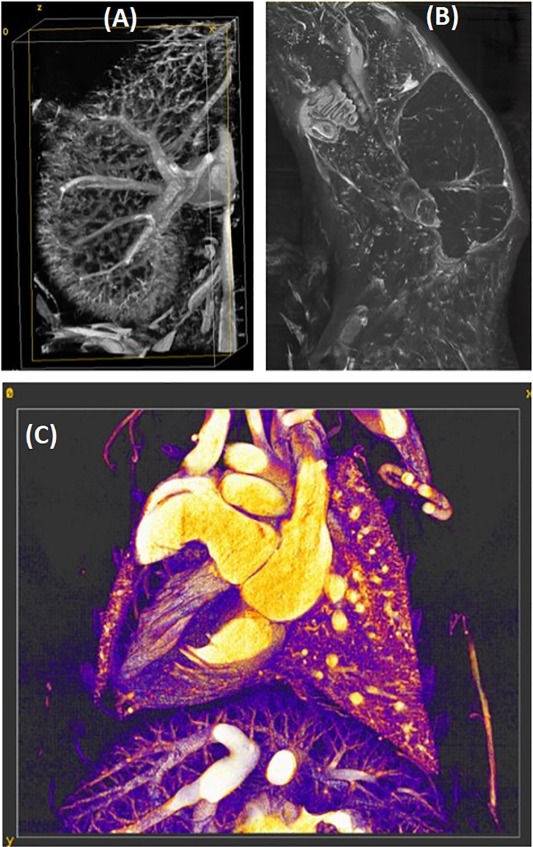
Examples of high-resolution, ex vivo vascular imaging using micro-CT and BriteVu as a vascular contrast agent. We illustrate mouse vasculature in the kidney (A), the head (B), and the thorax (C).
New deep learning paper on Clinical CT from our group: Evaluating renal lesions using deep-learning based extension of dual-energy FoV in dual-source CT—A retrospective pilot study.
Eur J Radiol. 2021 Jun;139:109734. doi: 10.1016/j.ejrad.2021.109734. Epub 2021

The code is available at: https://gitlab.oit.duke.edu/dpc18/duke-ct-spectral-extrapolation. It includes code for both of our DE extrapolation papers:
(1) Clark, D. P., Schwartz, F. R., Marin, D., Ramirez‐Giraldo, J. C., & Badea, C. T. (2020). Deep learning based spectral extrapolation for dual‐source, dual‐energy x‐ray computed tomography. Medical Physics, 47(9), 4150-4163.
(2) Schwartz, F. R., Clark, D. P., Ding, Y., Ramirez-Giraldo, J. C., Badea, C. T., & Marin, (2021). Evaluating renal lesions using deep-learning based extension of dual-energy FoV in dual-source CT – a retrospective pilot study. European Journal of Radiology, 109734.
Our QIAL papers presented at the SPIE Medical Imaging 2021:
Our keynote talk on Deep Learning Approaches in Spectral CT at the 2nd Annual Translational Imaging Conference AI and Machine Learning in Imaging.

We combined MRI and micro-CT to show that lack of GIT1 results in skull shape abnormalities, brain atrophy, white matter and cortical layer deficiencies. Clustering of volume covariance adjacency matrices identified vulnerable brain networks.

https://www.sciencedirect.com/science/article/pii/S0730725X20304537

