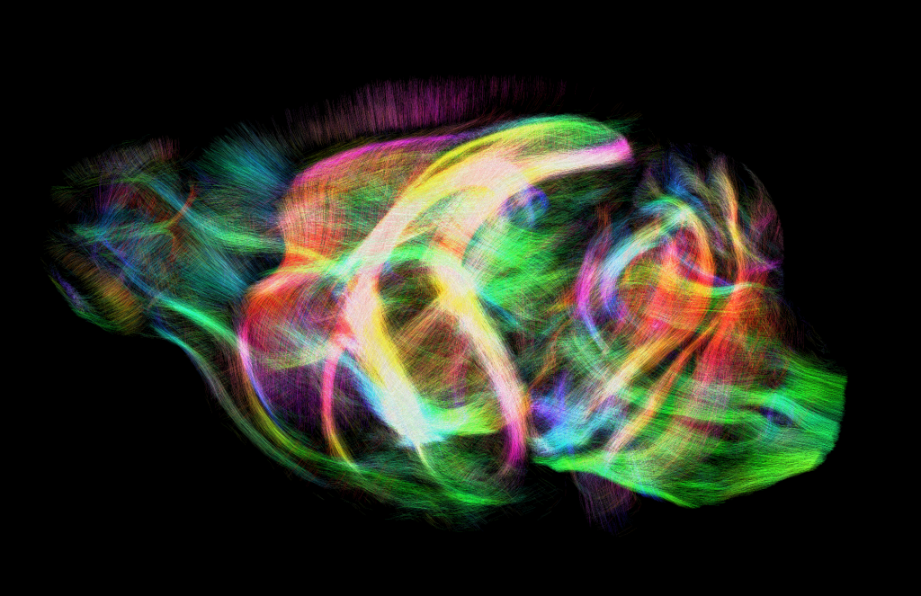
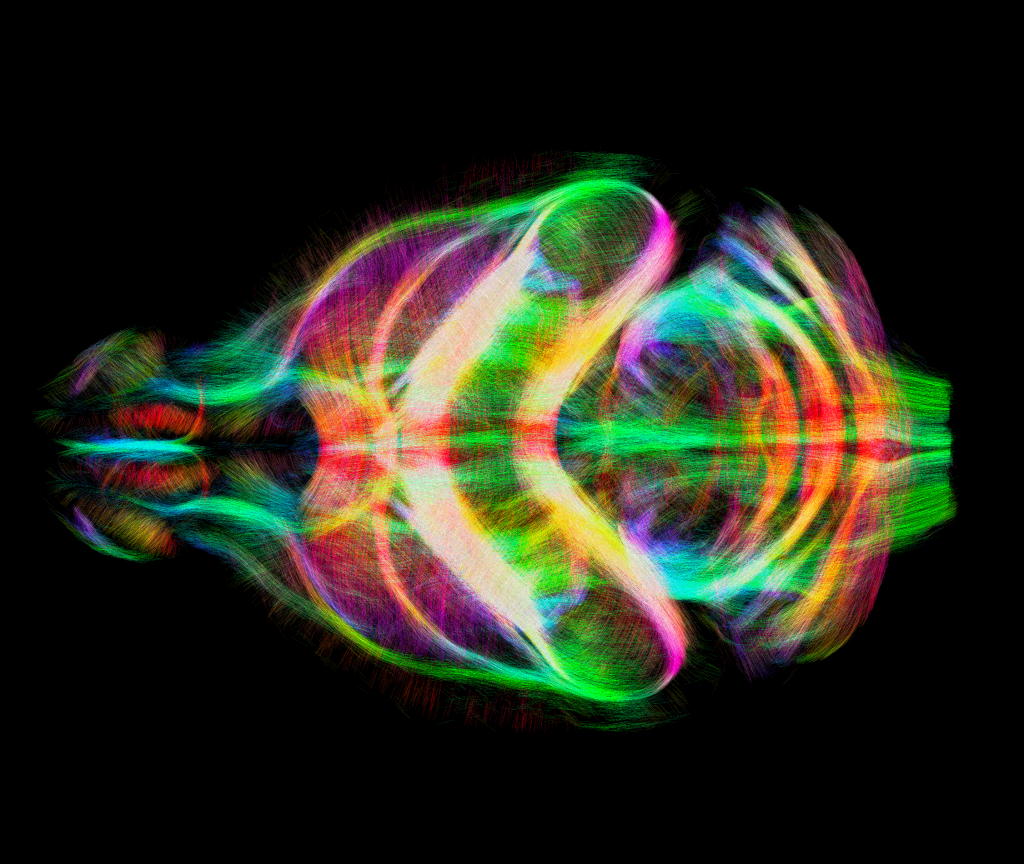
Tractography is a technique used to map neural connections in the brain using diffusion-weighted magnetic resonance imaging (DW-MRI). This technique involves measuring the diffusion of water molecules within the brain tissue and using this information to infer the orientation of axonal fibers, which make up neural connections between regions of the brain. By using specialized software to analyze the DW-MRI data, researchers can create three-dimensional maps of neural pathways in the brain, which can help them understand how different brain regions are connected and how information is transmitted between them. This technique has been used to study a variety of neurological conditions, such as Alzheimer’s disease, schizophrenia, and traumatic brain injury, as well as to investigate the neural basis of behavior and cognition.
Mouse tractography is a relatively new technique, but it has already proven to be a powerful tool for studying the mouse brain and could potentially have applications in understanding human brain connectivity and neurological disorders.
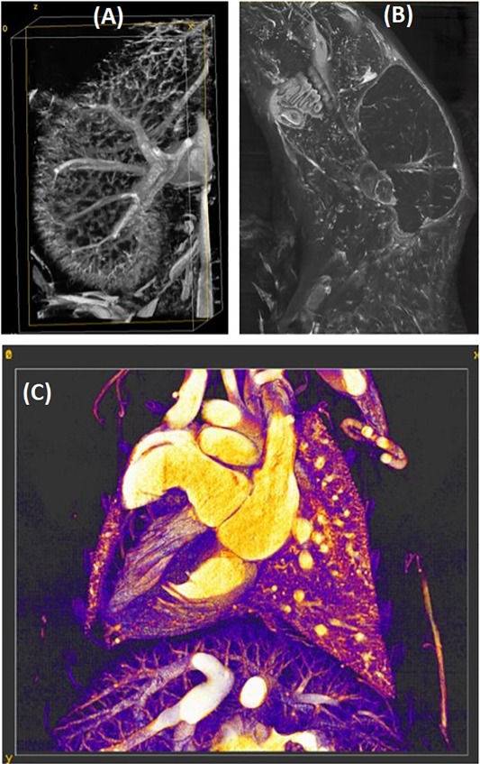
Using more than one b value or diffusion shells can help reduce the bias in estimating tissue diffusion properties because it allows for better characterization of the diffusion process in the tissue. The diffusion signal measured in MRI depends on the underlying microstructural properties of the tissue, such as the size, shape, and orientation of the diffusion barriers (e.g., cell membranes, axons, myelin). By acquiring diffusion-weighted images at multiple b values or with multiple diffusion shells, it is possible to obtain information about the diffusion process at different length scales and to disentangle the effects of different tissue compartments. For example, using two diffusion shells can help distinguish between intra- and extra-axonal diffusion in white matter. The first shell typically includes low b values, which are sensitive to the diffusion of water molecules within the axons (i.e., intra-axonal diffusion). The second shell includes higher b values, which are sensitive to diffusion outside the axons (i.e., extra-axonal diffusion). By fitting a model to the diffusion data acquired at both shells, it is possible to estimate parameters such as the axonal diameter, density, and orientation dispersion, which can provide insights into the microstructural properties of white matter.
49 direction MRI is a specific technique for performing DW-MRI in mouse tractography that involves acquiring images from 49 different directions. This allows for a more accurate reconstruction of neural connections in the brain, since it captures a greater range of diffusion orientations.
The following images are captured screens of 3D rendered brain tractography performed on a mouse: