Advanced Micro-CT and DSA Images Produced with Novel Instruments and Methods Developed at QIAL, Duke University
- Mouse leg with a sarcoma tumor imaged with high resolution micro-CT

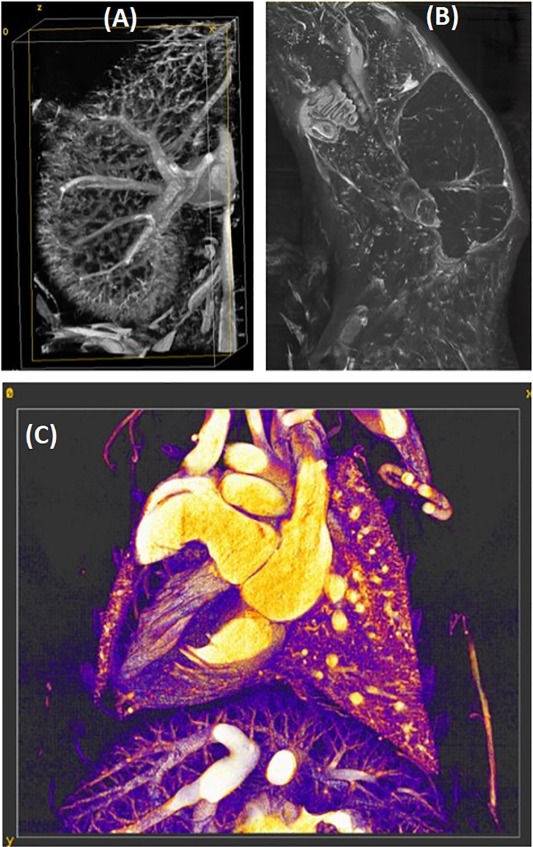
- Ex Vivo Vascular Micro-CT Images in Mice
-


- DSA of a tumor in a mouse leg

- DSA of a coronary arteries in a rat

- DSA of the cardiopulmonary system in a rat

- A rendering of the left ventricle of the beating mouse heart imaged with our Micro-CT

- Micro-CT of a femur in a rat

- Micro-CT of a 3D print of a mathematical object :

- A 3D rendering of a locust scanned with Micro-CT

Images generated with instruments and methods developed by the Quantitative Imaging and Analysis Lab (QIAL), Duke University.
Use of these images requires acknowledgment of QIAL and Duke University in any publications, presentations, or derivative works.