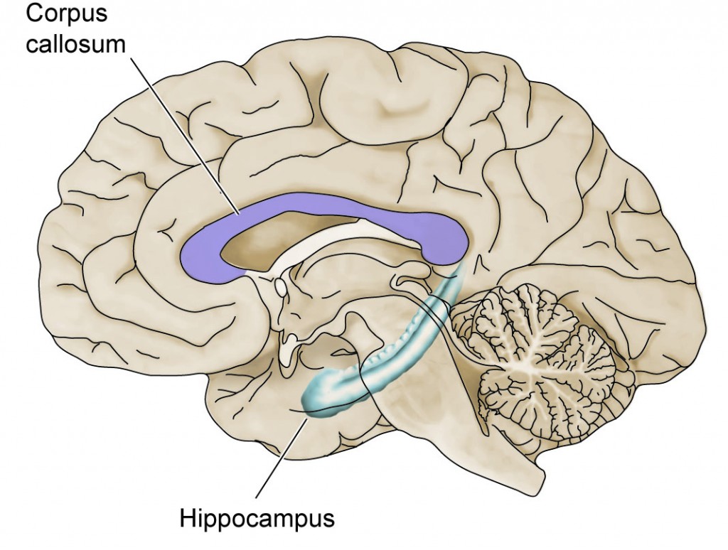
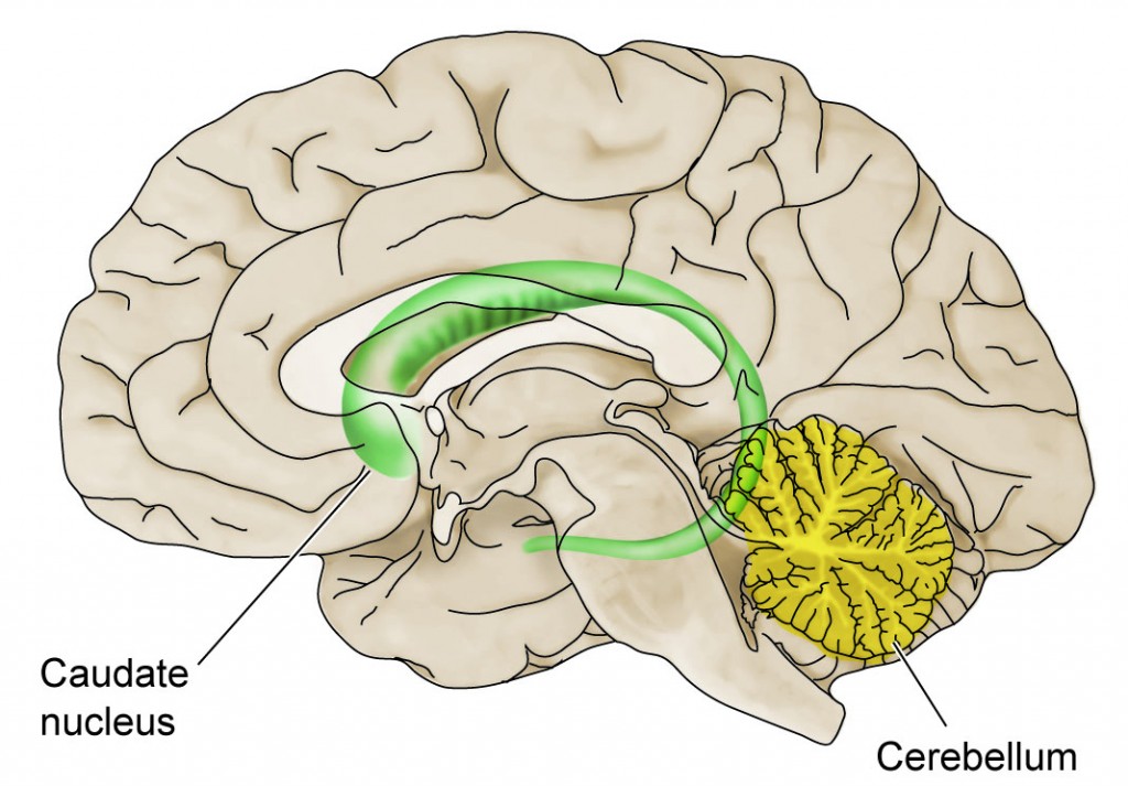
Sophisticated brain imaging technologies are now being used to identify the structural abnormalities present in the brains of living FASD children. Magnetic Resonance Imaging (MRI) has revealed an overall reduction in brain size, confirming previous autopsy findings. However, independent of the overall reduced brain size, MRI has revealed at least 4 major brain structures that are affected (i.e., reduction in size or abnormal shape) by prenatal alcohol exposure (Figure 4). These include: 1) the Corpus Callosum, a large bundle of nerve fibers that connects the two hemispheres together, enabling communication between the right and left brain; 2) the Caudate Nucleus, a structure that resides below the level of the cerebral cortex, which controls motor abilities and cognitive function; 3) the Hippocampus, another subcortical structure, which controls the ability to store new memories and participates in spatial learning; and 4) the Cerebellum, a structure that resides at the back of the brain, controlling motor skills, balance, and coordination. The importance of these brain regions in the neuropsychological and behavioral effects of prenatal alcohol exposure are discussed below.
Figure 4: Brain Structures Affected by Alcohol in Utero
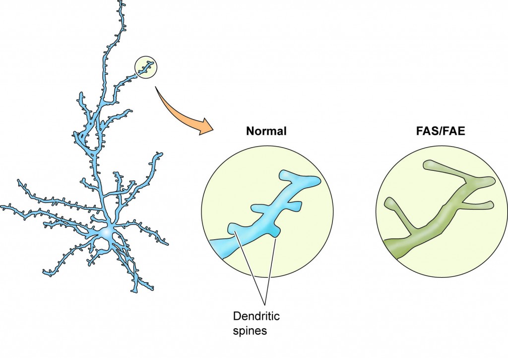
Using animal models (usually rats and monkeys) that mimic the effects of alcohol on human fetal brain development, scientists can demonstrate exactly how alcohol disrupts the development of these brain structures. For example, alcohol reduces the number of neurons within the cerebellum by suppressing cell proliferation and increasing neuronal apoptosis (genetically-programmed cell death; see above). Thus, alcohol triggers neuronal death during the 3rd trimester of otherwise healthy neurons. Alcohol also disrupts synaptogenesis or the proper formation of synapses—the connections between neurons that allow them to communicate. Animal models help scientists understand how this happens. When animals are exposed to alcohol in utero, the density of spines on the dendrites is reduced in areas such as the hippocampus, cerebral cortex, and cerebellum; the remaining spines become elongated (Figure 5), as if they are reaching out for communication with an axon terminal. Although it is difficult to obtain this information from children (via autopsy), there is one study that reports a greater than 50% reduction in dendritic spine density (and elongation of the spine) in a 4-month-old boy with FASD, compared to a healthy baby. The effect of alcohol on spine density and shape disrupts Synaptogenesis, ensuring that neurons will not function normally after birth. These events are particularly evident when binging takes place in the 3rd trimester.
Figure 5: Loss of Dendritic Spines After Exposure to Alcohol in Utero
In addition to imaging structural abnormalities, other forms of brain imaging can reveal functional abnormalities within specific brain areas. For example, scientists have shown that FASD children have a delay in a certain type of electrical activity within the Parietal Cortex that is associated with information-processing. In addition, imaging techniques, such as Positron Emission Tomography (PET), can be used to study the metabolic activity of the brain (i.e., how well it is actually working). In FASD children, there is a reduction in metabolic function within the caudate nucleus as demonstrated by PET imaging. When combined with structural information from MRI studies, these studies can provide a powerful picture of the damage to specific regions within the working brain.