In the past, scientists theorized that the neuropsychological effects in FASD children could be attributed to damage of specific brain structures. Now by using MRI to see the damage in living subjects, researchers are performing studies to link specific brain region structural damage with specific neuropsychological deficits in FASD children. A review of the areas most affected by prenatal alcohol exposure and the corresponding neuropsychological problems follows below.
Cerebral Cortex
The Cerebral Cortex (the outer part of the brain, see Figure at right) is responsible for several functions that involve higher levels of control and organization. Such functions include: sensory and motor control, Cognition and abstract thought, working memory, speech and language, and visual and hearing perception. Executive functioning, which is dependent on working memory, is associated with the cortical areas in the frontal lobe (the Prefrontal Cortex). Much of the final circuitry within the cerebral cortex is established during the 3rd trimester, so any or all of these cortical functions can be disrupted if the fetus is exposed to alcohol during this time.
Cerebellum
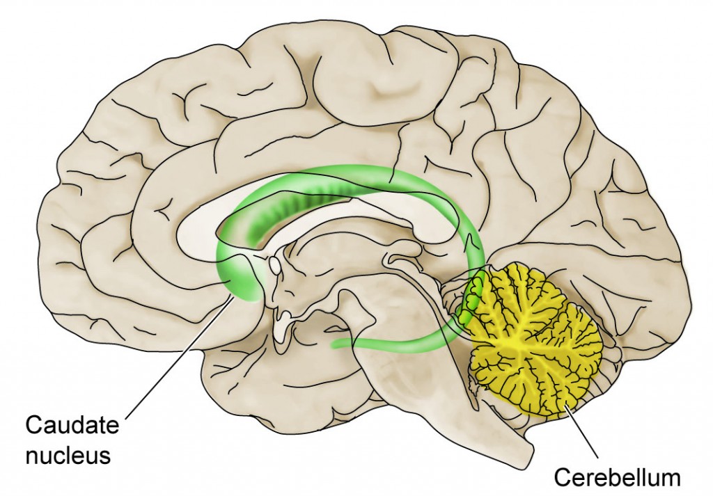 The cerebellum (in yellow) is a structure that has not completed its development at birth; it continues to form its circuitry early in the postnatal period. If the fetus is exposed to alcohol as late as the 3rd trimester , there can be considerable effects on cerebellar function. In addition to the motor control and coordination provided by the cerebellum there are several other functions. It helps to provide cognitive processing, acquisition of language fluency, task sequencing, and time perception and estimation. Children with FASD are dysfunctional in each of these tasks.
Caudate Nucleus
The caudate nucleus is part of the Basal Ganglia. It, too, plays an important role in motor function. However, the caudate is also important in cognitive function, motivation, and executive functioning, or the ability to plan and execute specific tasks. FASD children have particular trouble with executive function. The basal ganglia work together with the cerebellum in cognitive and attention tasks; damage to the caudate by in utero alcohol can be expected to result in attention deficit problems manifested in the classroom.
Corpus Callosum
One of the most consistent defects in brain structure in people with FASD is the reduction in size, altered shape, or complete absence of the corpus callosum. Examples of MRIs of a normal child and an FAS child with a missing corpus callosum are shown in Figure 7.
Figure 7: MRI Images of Normal and Abnormal Brain Structure

This major bundle of nerve fibers that connect the left and right brain is important in timing tasks, motor tasks, and coordination. Failure of the corpus callosum to develop properly leads to slow reaction times and anything from mild cognitive impairment to extensive mental retardation. In an MRI study of children and young adults with FASD, the damage to the corpus callosum was associated specifically with verbal learning ability. In another study of adults with FASD, different corpus callosum shapes were associated with deficits in Executive Function and motor function; it did not matter if there were facial dysmorphologies. Although the corpus callosum is formed around the 10th week of gestation, it undergoes a major growth spurt relatively late in pregnancy (at about month 6 or 7). Drinking during the 1st trimester can prevent its formation; drinking during the 2nd or 3rd trimester can affect its shape and ability to communicate with neurons in cortical areas.
Hippocampus
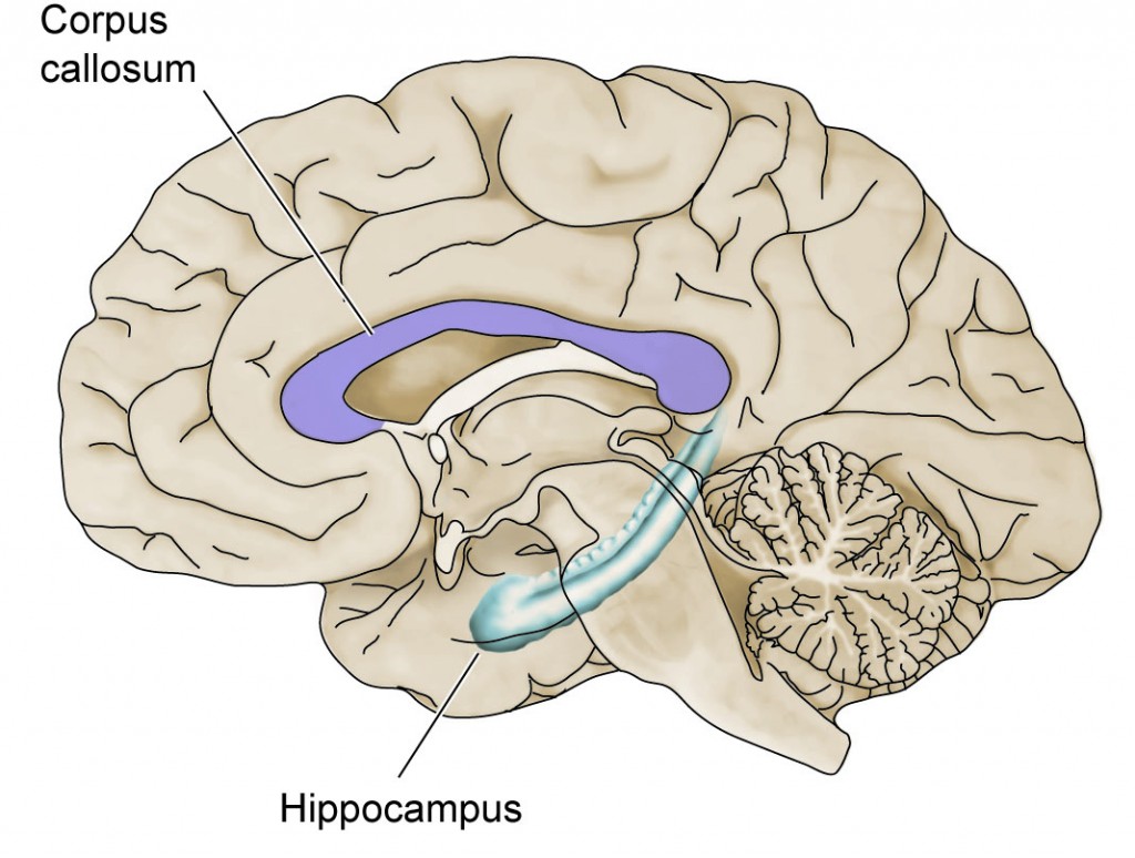 The major structure in the brain responsible for learning and memory is the hippocampus, although other structures such as the cerebral cortex are involved as well. The circuitry of the hippocampus is established rather late during pregnancy and exposure of the fetus to alcohol in the 3rd trimester (or before) can be expected to have significant effects on learning and memory.