The brain develops throughout pregnancy, and continues developing even through adolescence. During the first 10 weeks of pregnancy, the basic cells that make up the brain are formed (Figure 2). These include neurons, which eventually send electrical and chemical signals to one another, and glial cells, which provide both structural and chemical support to the neurons. During the 1st trimester, the neurons and glia undergo cell division, multiplying aggressively in preparation for their subsequent organization into specialized functions.
Figure 2: Stages of Brain Development Throughout Pregnancy
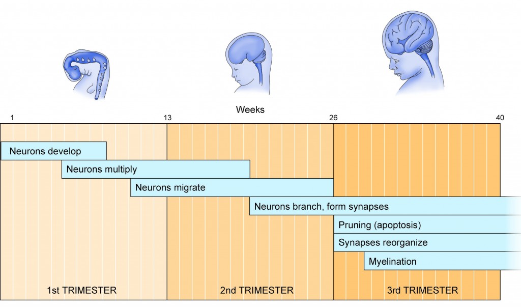
As pregnancy moves into the 2nd and 3rd trimesters, several events take place. The neurons grow, forming numerous branches (Dendrites) and an axon, structures that are crucial in sending and receiving information (Figure 3). The dendrites grow small protrusions called Spines—this increases the surface area for communication between neurons (see below). At the same time, the axons become surrounded by myelin, a fatty sheath provided by glial cells, which will help the neurons conduct electrical impulses over long distances.
Figure 3: Neuron Structure

A typical neuron is shown making a synapse with another neuron. The cell body contains the nucleus where the DNA resides. A long axon leaves the cell body and ends in a terminal. The terminal makes a connection (synapse) with the dendrites of another neuron. An enlarged view of a synapse is shown at the bottom. Within the synapse, neurotransmitters are released from the terminal of a neighboring neuron; the neurotransmitters bind to specific receptors located on the dendritic spines of the receiving neuron. Receptor binding leads to either electrical or chemical signals in the receiving cell.
Once the structural aspects of neuronal development are in place, several chemical events emerge. The neurons start to synthesize their own chemicals, called Neurotransmitters. Examples include Dopamine, Serotonin, Norepinephrine, Glutamate, and γ-Aminobutyric acid (GABA). In the postnatal brain, these neurotransmitters signal neurons to perform work. The neurotransmitters are released from the terminals of axons in response to electrical signals, and they bind to special proteins called Receptors on a neighboring neuron (Figure 3).
There is a high density of these receptors on the Dendritic Spines of neurons. Each neurotransmitter binds to its own receptor; serotonin binds to serotonin receptors; and glutamate binds to glutamate receptors. All of this activity takes place in the Synapse or the connection between two neurons. The consequence is a change in the rate at which the receiving neuron conducts an electrical impulse. It is the rate at which neurons fire impulses within different brain regions that underlies every function in the brain, whether it is motor control, speech, learning, attention, or judgment.
However, in the developing brain, this sophisticated function of neurotransmitters is not yet needed. Instead, the neurotransmitters play a different role; they serve as growth factors,directing neurons to establish connections (i.e., synapses) with appropriate neighbors bearing the corresponding receptors that will be needed for future communication.The instructions provided by the neurotransmitters and their corresponding receptors during development are crucial to the formation of functional and efficient synapses that are needed after birth.
Once the major neuronal connections are formed, there is some “pruning” that must be performed. This pruning is called Apoptosis, a genetically-programmed form of cell death. Apoptosis helps eliminate those neurons that don’t grow very well during the first two trimesters—there aren’t quite enough growth signals provided by neurotransmitters and other growth factors to reach every neuron. Thus, apoptosis ensures that any neurons that don’t grow properly are eliminated so that neuronal transmission will occur normally. Apoptosis continues well into post-natal development.
All of these developmental events progress at different rates within different brain regions. Such variation in developmental rates may explain, in part, the differential effects of alcohol, depending on when it is consumed during pregnancy.