
Meniscus Injuries
Cartilage Strain After Meniscus Injury
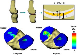
Strain (ε) across the tibiofemoral joint was approximated from the overlap in cartilage surfaces. The penetration distance (d) was divided by the combined thickness of the undeformed tibial (t1) and femoral (t2) cartilage surfaces at each point within the joint to create a strain map (top). Contact strain maps were created for all angles of both the injured and uninjured knees. The strain maps are shown for one subject at 601 of flexion. With a meniscus tear (bottom right), the cartilage contact strain increased in the medial compartment, as compared to the uninjured knee (bottom left).
Meniscus Coverage Impacts Tibial Cartilage Deformations
 Representative color thickness maps of tibial cartilage before and after activity. Thicker cartilage is indicated by red, and thinner cartilage is indicated by blue. The meniscal footprint is outlined in black.