Home » Uncategorized (Page 2)
Category Archives: Uncategorized
Our PLOS ONE paper is now published!
Bridging the translational gap: Implementation of multimodal small animal imaging strategies for tumor burden assessment in a co-clinical trial
S. J. Blocker, Y. M. Mowery, M. D. Holbrook, Y. Qi, D. G. Kirsch, G. A. Johnson, C. T. Badea, PLoS ONE 14(4): e0207555. (2019)

U24 DICOM tool
Download our U24 DICOM tool
This is a MATLAB-based tool for converting 3D CT image volumes from NifTi format to DICOMs. This repository contains a GUI which can be used to assign values to common DICOM fields. It also contains functions which can be used in your own code to streamline workflows.

Duke Standard Operating Procedure for MR Imaging at 7T of tumor-bearing mice using a surface coil
Generalized Scanning Protocol (U24) – SOP

This SOP is designed as a general guide for in vivo MR imaging of small animals as part of a pre-clinical cancer study, using a volume transmit coil + surface receive coil at high fields. Note that this protocol begins with information that is not machine or project specific.
SPIE manuscript on the importance of gating for preclinical CT imaging of lung nodules
S. J. Blocker, M. Holbrook, Y. M. Mowery, C. T. Badea. To gate or not to gate: an evaluation of respiratory gating techniques to improve volume measurement of murine lung tumors in micro-CT imaging. Proceedings Volume 10953, Medical Imaging 2019: Biomedical Applications in Molecular, Structural, and Functional Imaging; 109531H (2019) https://doi.org/10.1117/12.2512534
Event: SPIE Medical Imaging, 2019, San Diego, California, United States

SPIE manuscript on sarcoma segmentation
M Holbrook, SJ Blocker, YM Mowery, CT Badea. Multi-modal MRI segmentation of sarcoma tumors using convolutional neural networks.
Proceedings Volume 10948, Medical Imaging 2019: Physics of Medical Imaging;
Event: SPIE Medical Imaging, 2019, San Diego, California, United States


Welcome to Duke Preclinical QIBA!
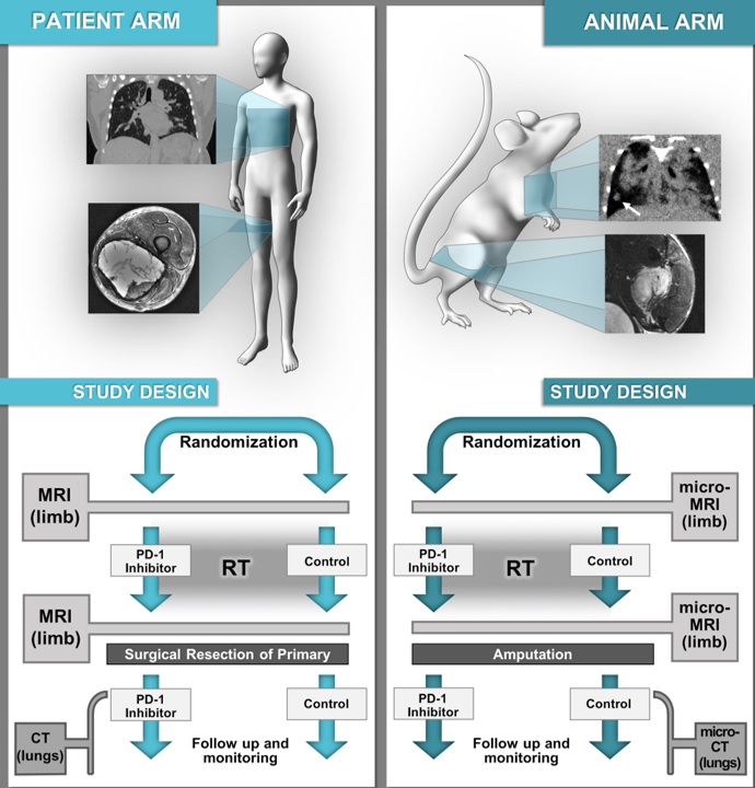
We are members of the NIH Co-Clinical Imaging Research Resources Program (CIRP). CIRP promotes development of quantitative imaging resources for therapeutic or prevention co-clinical trials that study both patients and human-in-mouse models.
Our project entitled “The Duke Preclinical Research Resources for Quantitative Imaging Biomarkers,” focuses on optimizing preclinical MRI of the extremity and respiratory-gated CT of the lungs for utilization in a co-clinical trial evaluating radiation therapy and immunotherapy with a PD-1 inhibitor in a genetically engineered mouse model (GEMM) of soft tissue sarcoma. We make available:
Recent Comments