Human brain development begins shortly after conception and continues through adolescence until adulthood. During the first 10 weeks of pregnancy (the first trimester), a group of cells in the embryo called stem cells receive chemical signals to develop into the primary cells of the brain, called neurons and glia. The birth of new neurons and glia is referred to as neurogenesis and gliogenesis, respectively. Ultimately, neurons will be responsible for communicating messages throughout the entire brain and the glial cells will provide both structural and chemical support to the neurons.
Although most of the neurogenesis occurs in the fetus and newborn, the “birth” of new neurons actually continues in some parts of the brain right through old age! The fetal brain contains as many as 100 billion neurons at birth.
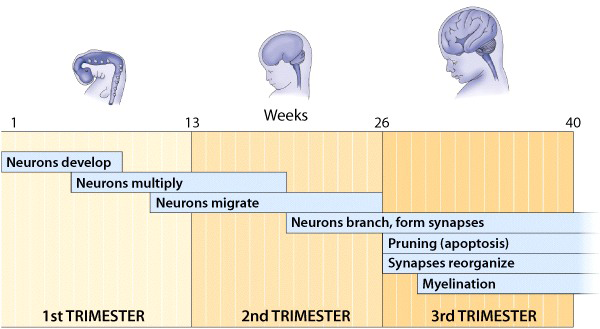
As neurons develop, they receive “cues” from their surrounding environment (such as neurochemicals and proteins in the vicinity) that direct them to migrate to their final destination within the brain. Organization of the brain is dependent on the migration of neurons to the appropriate position within the brain.
As pregnancy moves into the 2nd and 3rd trimesters fetal precursor neurons continue to multiply and differentiate into mature neurons that migrate to their designated area. As neurons mature in structure, they form numerous branches (dendrites) and an axon, structures that are crucial for receiving and sending information, respectively. At the same time, some axons become surrounded by myelin, a fatty sheath provided by glial cells, which will help the neurons conduct electrical impulses over long distances.
Once neuronal structures are in place, several chemical events begin. The neurons start to synthesize their own neurotransmitters, the chemicals responsible for communication between neurons.
Review neuron structure and function
However, in the fetal brain, this sophisticated function of neurotransmitters is not yet needed. Instead, neurotransmitters play a different role; they serve as growth factors, directing neurons to establish connections (i.e., synapses) that will be needed for future communication. The instructions provided by the neurotransmitters and their corresponding receptors during development are crucial to the formation of functional and efficient synapses.
By the third trimester and after birth, the brain directs the elimination of unnecessary neurons. The loss of neurons is a part of a normal “pruning” process. Neurons that make inappropriate or redundant connections are selectively eliminated by a process called apoptosis. Apoptosis is a form of cell death that proceeds by a series of cellular reactions under control of the cell’s genetic instructions. Those neurons that are not eliminated by apoptosis continue to grow, establish new connections with other neurons, and strengthen the connections already in place.
All of these developmental events progress at different rates within different brain regions. Such variation in developmental rates may explain, in part, the different effects of chemicals such as alcohol on the fetal brain depending on when during pregnancy they are consumed.
Learn more about apoptosis