While prenatal alcohol exposure affects every area within the fetal brain, some areas are exquisitely sensitive to the damage by alcohol. In the past, scientists hypothesized that the cognitive and behavioral problems of children with FASD could be attributed to the damage of specific brain structures. More recently, scientists have used newer technologies to image the brain, both in humans and in animal models. Now researchers can actually see brain damage in living patients and determine how damage to certain brain regions causes specific cognitive problems.
The brain regions most affected by prenatal alcohol exposure that have been linked to cognitive and behavioral problems are reviewed below.
Cerebral cortex
The cerebral cortex (the outer part of the brain) is responsible for several functions that involve attention, memory, perceptual awareness, thought, language, and consciousness. Executive function, which includes planning, self-control, and abstract thinking, is associated with the part of the cortex in the front of the brain (the “prefrontal cortex“). Many of the final connections of neurons within the cerebral cortex are established during the third trimester (and even during childhood and adolescence). Any or all of these cortical functions can be disrupted if the fetus is exposed to alcohol during this time.
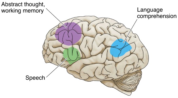 Figure 5.8 Areas in the cerebral cortex that regulate memory, language comprehension, and speech can all be affected in a fetus when a mother drinks alcohol.
Corpus callosum
One of the most consistent structural brain defects in people with FASD is the malformation or absence of the corpus callosum, which is a bundle of nerve fibers that cross from one side of the brain to the other, connecting the left and right hemispheres. The corpus callosum can be seen easily in a magnetic resonance image (MRI) of the midline of the brain in a side view. Look at the MRIs of the corpus callosum in the normal adolescent and in an adolescent with FASD (who is missing a corpus callosum) (Figure 5.9)
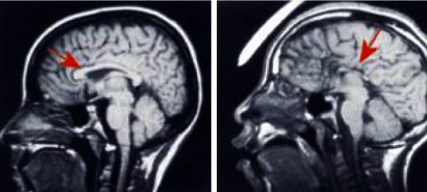 Figure 5.9 On the left is a magnetic resonance image (MRI) of the brain of a normal adolescent (age 14). The red arrow points to the corpus callosum, the white bundle of fibers that crosses over the midline. The MRI on the right is of an adolescent with FAS (age 14); the red arrow shows the lack of a corpus callosum. [MRI photos courtesy of Drs. Ed Riley and Sarah Mattson, San Diego State University]
Learn more about MRI
The corpus callosum is important in timing tasks, attention, motor tasks, and coordination. When the corpus callosum fails to develop properly a person tends to have problems with attention needed to perform cognitive (thinking) tasks, poor motor coordination, and even mental retardation. Importantly, these defects in brain function can occur even when facial abnormalities are not present. Because the corpus callosum develops throughout gestation, it is sensitive to alcohol exposure during all stages of pregnancy.
 Figure 5.10 Prenatal alcohol exposure can damage the corpus callosum and hippocampus, causing cognitive, attention, and memory problems.
Children with FASD typically have difficulty thinking through things (and poor attention) and some children also have problems with motor coordination.
Hippocampus
The major structure in the brain responsible for learning and memory is the hippocampus, although other structures such as the cerebral cortex are involved as well. The formation of the hippocampus begins early in the second trimester and the proper connections of neurons in the hippocampus are established rather late during pregnancy. Exposure to alcohol at any time during pregnancy can alter hippocampal formation. The consequences of hippocampal damage are learning and memory problems.
It is very common to find children with FASD who have short term memory and learning problems.
Learn more about memory
Caudate nucleus
The caudate nucleus plays an important role in both understanding (cognition) and movement. However, the caudate is also important in the ability to plan and carry out specific tasks. Children with FASD have particular trouble with such “executive” functions. The caudate works together with the cerebellum (see below) in cognitive and attention tasks; alcohol damage to the caudate of a developing brain can result in problems with attention.
Attention deficits are very common in children with FASD.
 Figure 5.11 Prenatal alcohol can cause damage to the caudate nucleus and cerebellum, causing problems with attention and cognition.
Cerebellum
The cerebellum, which is responsible for motor control and coordination, is a structure that has not completed its development at birth; it continues to form its circuitry early in the postnatal period. In addition to motor control and coordination, there are several other functions of the cerebellum. It helps with cognitive processing, acquisition of language fluency, and time perception and estimation.
Children exposed to alcohol during gestation can have problems with motor skills, cognition, language skills, and ability to assess time.
Summary of brain regions most affected in FASD
| Brain Region cerebral cortex corpus callosum hippocampus caudate nucleus cerebellum |
Function affected by alcohol in utero executive function, attention, memory, language motor tasks, coordination, attention learning and memory cognition, movement, attention motor control, coordination, and time perception |