Tag Archives: RF2022-Week2

CRISPR-Cas9 and Parkinson’s Disease

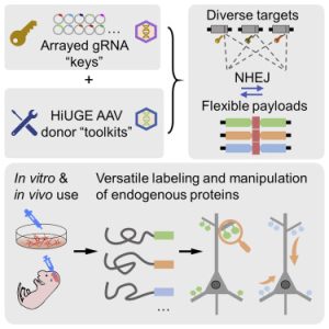
One of the focuses of the Soderling Lab is the mechanisms underlying disrupted synaptic connections, abnormalities that play a role in the onset of neurodegenerative and psychiatric disorders. To accomplish this, the lab has developed a combination of proteomic and CRISPR approaches, such as the CRISPR-Cas9-based Homology-independent Universal Genome Engineering (HiUGE) method. HiUGE allows efficient and convenient alteration of endogenous proteins through adeno-associated virus (AAV) vectors of insertional DNA sequences that can merge into the part of the genome specified by guide-RNA vectors (Gao et al., 2019). As demonstrated in the following Graphical Abstract from Gao et al., HiUGE provides a simplified way to modify proteins in vivo and in vitro for the study of gene and proteins functions, thus making it easier to investigate the proteins and synaptic disruptions involved in diseases such as Parkinson’s (2019).


Parkinson’s disease (PD) is a progressive, age-associated neurodegenerative movement disorder often characterized by a resting tremor, slow movement, and difficulty maintaining posture due to the loss of dopamine-producing neurons in the midbrain. The etiology of the disease continues to be studied, with advances in the identification of gene mutations specific to familial PD encouraging further inquiry into the mechanisms by which these mutations induce it. One such PD-linked gene is the vacuolar protein sorting 35 ortholog (VPS35), with the VPS35 D620N mutation having been identified as pathogenic (Williams et al., 2017).

Together, HiUGE and PD are the foundation for my research project this summer. I will be investigating the factors that may improve the efficacy of the former while simultaneously employing HiUGE to observe the latter in vitro (and possibly in vivo). Currently, I have three research questions that include the following:

  1. Are dual-oriented HiUGE-donors more efficient than single-oriented ones for gene expression?
  2. What is the effect of knock in WT/DG20N VPS35 – a dual-oriented HiUGE-donor – in vitro (and potentially in vivo)?
  3. Does UltraID produce biotinylation of alpha synuclein comparable to TurboID?

The first question relates to whether a DNA vector or construct created through the HiUGE method might produce double the expression of a specific gene if its epitope is dual-oriented versus single-oriented. The second question involves a dual-oriented HiUGE donor with healthy and PD-linked DNA fragments being introduced in vitro to a cell culture and potentially in vivo with mouse models. This second question also involves partially testing the efficacy of the dual-oriented HiUGE donor. Lastly, the third question has ramifications as to whether labeling proteins such as alpha synuclein and others potentially linked to PD in the synapses can be equally if not more effective through the use of the enzyme UltraID, a smaller molecule as opposed to the currently used TurboID.

I look forward to continuing to learn as I seek the answers to these and other questions in the Soderling Lab this summer.

Pain, Pain, Pain

Second week of BSURF done and honestly, I am not 100% sure what it is that my summer project consists of yet. I know the general gist of what it is and the purpose of doing it so here goes my little elevator speech about it: Our goal is to study a protein that has been found to alleviate neuropathy induced pain when administered in animal models more in depth. Neuropathy refers to nerve damage usually caused by disease or injury. Chronic nerve pain is a problem because common medicine like Ibuprofen, Advil, or even opioids are not effective in this case as they work in a different manner that does not target the root problems of nerve pain. 

Immunohistochemistry (IHC) will be used extensively in order to visualize our protein of interest’s receptors within DRG neurons. To do this, the slide with the tissues of interest is prepared with a washing buffer first, then stained with a primary antibody which is what binds to the receptor of interest. The slides are left in the cold room overnight and the following day they are again stained with a secondary antibody which binds to the primary antibody that was previously added. It is this secondary antibody that becomes fluorescent under the microscope and allows us to see if and where our receptors of interest are in the samples we are looking at. 

This will be done with the hopes of better understanding how our protein of interest works when said protein is administered to an organism and alleviates pain to determine if it could potentially be applied the same way in humans. In preparation to carry out this project, I have been receiving training in the proper handling of the animals in our lab, how to do behavior tests, prepare slides with tissue samples, and learning how to do the IHC protocol needed. I’m very excited to see how this project evolves and see the data we collect in the following weeks.

Picturing the Brain

Visualizations are a key tool for researchers to be able to communicate their data and results. Great visuals can give scientists the power to internalize complex systems. This summer, I am working with Dr. John Pearson’s lab on their improv software, which has its own visualization of real-time neural activity when implemented with neurobiology labs. For my research project this summer, I am creating a new visualization that integrates seamlessly with the improv software and improves upon the legacy visualization.

Currently, Anne Draelos, a postdoc at the Pearson Lab, collaborates with Dr. Eva Naumann’s lab to implement improv in their experiments with zebrafish. The zebrafish are shown various stimuli to mimic shadows that would be seen in the water, and improv identifies real-time neural activity that correlates to each stimuli. After a few rounds of different stimuli, improv then predicts and suggests which stimulus will prompt the most neural activity. During this entire process, improv displays its GUI (a graphical user interface, also referred to as “gooey”). This GUI contains a live image of active neurons that are color coded for their correlating stimuli, a line graph of population neural activity, and a replica of stimuli being shown. Below is an example of the GUI in action from the improv paper.

For my new visualization, I plan to improve on the legacy GUI by making it more accessible to users through a platform called Jupyter Notebook. This can be accessed through a browser, unlike the old PyQt platform used. I also plan to add a wider variety of graphs and plots to aid experimentalists’ understanding of the data. Some examples include histograms, scatter plots, and video plots of the current stimuli and what improv suggests. My project will be split into two major steps:

  1. First, I will be exploring how to create plots with live updates of the data. This will require me to learn how to use Jupyter Notebook as my base platform and learn how to plot some basic data. Then, I will explore some Python packages to help with the live aspect of the data. The incoming data needs to be integrated into existing plots in a fast and efficient way.
  2. Next, I will look into the best ways to access the actual data from improv and incorporate it into the Jupyter Notebook. One potential solution is to implement plasma, a software developed by Apache Arrow.

Overall, I am excited to tackle this project and contribute to the future implementation of improv!

Actually Getting Started

Dr. Huang’s lab focuses on the study of ovarian cancer. She is specifically interested in studying the tumor microenvironment, or how the environment around a tumor affects the way it functions. My project will focus on the relationship between age and how receptive cancer cell lines are to chemotherapy. Previous studies have shown that older patients suffering from ovarian cancer have significantly worse outcomes than those that are younger. My project seeks to understand some of the mechanisms that may lead to the worse outcomes among older patients.

In all honesty, I don’t have all of the details about my project ironed out just yet because my professor has been out of the lab for the last two weeks on a previously planned vacation. But, she gave me a basic outline of my project and some literature to look at before I got the chance to get started working on it when she’s back. I’ll be culturing a cancer cell line, HEYA8, to look at the effects of aging on ovarian cancer. These cancer cells will be cultured in the medium from preadipocytes. Preadipocytes are fat precursor cells and they are part of the tumor microenvironment for ovarian cancer tumors. I’ll end up comparing how sensitive these ovarian cancer cells are to chemotherapy depending on whether they were grown in medium from aged vs. young preadipocytes. I will also be culturing the preadipocytes myself, and they will be cultured at different doubling times in order to create the aged vs. young lines. I’ll be conducting chemosensitivity tests on the cancer cell lines in order to evaluate if the age of the preadipocytes makes a difference.

The Great Filter of Metastasis: Where is it?

Metastatic cancer is what is often referred to as stage 4 cancer, and describes the development of cancer where the cancerous cells spread to other organs in the body. This process is called metastasis. Metastasis is an important occurrence that often determines the fatality of cancer for many people, making it a significant point of research worldwide. Metastasis is considered a multi-step cascade involving the following key processes (Lambert et al., 2017):

  1. Intravasation: Cancerous cells leave the primary site (original tumor) and enter the bloodstream
  2. Survival in circulation: The cells survive while moving through the body in the bloodstream
  3. Extravasation: The cells manage to exit the bloodstream and breach the extracellular matrix of other organs
  4. Engraftment: Cancer cells adhere to the tissues of foreign organs
  5. Metastatic Colonization: Now-metastatic cells proliferate and form cancerous lesions in the foreign organs creating secondary tumors

Y. J. Tang et al., 2019 with the Alman Lab at Duke conducted a study with undifferentiated pleomorphic sarcoma (UPS) in mouse models that indicated that although a variety of clones (i.e. cancer cell types) completed the intravasation and survival in circulation steps of metastasis, only a single clonal variant was responsible for the development of secondary lesions in the lung. The term metastasis-initiating cells (MIC) refers to these cells that can survive and form secondary lesions. The survival of solely MICs means there is something, some physical or chemical wall, a filter of sorts, that weeds out the rest of the cells such that only a select type can survive and proliferate in the lung.

Graphical Abstract from Y.J. Tang et al., 2019 where you can see that there is a heterogenous mix of cell types in the primary tumor, but a single type in advanced metastasis.

This summer I am working to investigate what this filter might be. Using a microfluidic device, or a small device that has microchannels and chambers which cells can be seeded into, I will be studying the physical properties of MICs. One of the first things I will be considering is the process of extravasation, where MICs must travel through microbial spaces in the extracellular matrix. Using a microfluidic device with varying dimensions of microchannels, hopefully, I might be able to identify both the deformability of UPS MICs (how much they can deform to squeeze through a small gap) and if only MICs are able to deform to get through certain size channel. This may demonstrate a potential point in metastasis where the filter may lie.

Continue reading

Yersinia pestis and human genetics

The Ko lab studies how genotypic variations contribute to the severity of infectious diseases. By infecting almost a thousand different cell lines, the lab has identified a single-nucleotide polymorphism (SNP) associated with variations in infection phenotypes by Yersinia pestis, the causative agent of the bubonic plague. The SNP is on a gene that codes for a transmembrane immunoreceptor. My mentor Rachel’s project focuses on the specific interactions between Y. pestis and the protein product of this gene. One way of confirming the role of the protein in bacterial invasion is to see if the clustering of this protein around Y. pestis changes for cells overexpressing mutated versions of this gene. The extracellular portion of this protein contains several immunoglobulin (Ig)-like domains. Based on the preliminary data from an experiment comparing all proteins in the same family as the protein of interest, Rachel suspects that Ig-like domains 1, 2, and 3 affect bacterial attachment. 

My project for the summer is to help design and build mutants that exclude each of the three domains. Infection of cells expressing the mutants can then show whether and how these Ig-like domains affect bacterial attachment and invasion. To accomplish this goal, I’ll first remove the DNA sequence coding for Ig-like domains 1, 2, and 3 from a plasmid that codes for the protein of interest. I’ll then ligate, separately, three mutated DNA fragments into the plasmid. HeLa cells transfected with the new plasmids will then express the mutated versions. Finally, infecting these HeLa cells with Y.pestis will reveal the critical domains for bacterial ligand binding. This project will contribute to the overarching goal of understanding how Y. pestis utilizes this protein to accomplish cellular invasion and how different genotypes of the protein affect Y. pestis infection.

Comparing Creeks: Bugs as Bioindicators

This summer, I am working with the Bernhardt Lab, a lab focused on aquaterrestrial biogeochemistry. Essentially, this entails ecology, ecosystems, and ecotoxicology. My contribution to the lab is helping sample, count, and identify aquatic macroinvertebrates in local streams, creeks, and watersheds as bioindicators for water quality and stressors (like pollutants) present at different sites.

My project specifically concerns three different creeks, one of which is largely forested with minimal pollution, and the other two are more urbanized and polluted. Of these latter two, one is located before a waste water treatment plant, and the other is placed after the waste water treatment plant empties out its treated water. I will be counting and identifying aquatic insects from each of these sites with samples collected from the exact same area over all four seasons. The goal of my research is to identify insects from each of these different sites while also comparing which insects are present across the seasons.

Of course, there are limitations to this research. This is a descriptive study which relies more heavily on relativity in respect to reproducibility. The sampling and subsampling methods will be consistent so that I will be able to compare my results within the scope of my own research. In representing my data, I hope to analyze the number of insects present at each site across seasons, as well as their body size/density and biodiversity.

I am very excited for my project, as bugs can tell us a lot about the types of stressors present at different sites, especially ones so localized to one another. The health of our environment and the ecosystems around us are of the utmost importance, and I am very glad to be contributing to research efforts to improve this.

Lions, Fibers, and Bacteria, Oh My!

The scene opens up to a birds-eye view of the African savannah. From afar, all seems calm: the tall grass sways in the wind as sunlight bathes the sparse trees and bushes in gold. But as you zoom in closer there’s something else: a cluster of dark shapes in motion on the horizon. Sounds of a feeding frenzy hit you before you can distinguish one animal from another in the mass: the yapping of hyenas and screeching of vultures as they descend on an fresh antelope carcass. A pride of lions pads away, licking their jowls and letting the scavengers finish the rest.

This is our classic idea of an ecosystem. It is also one of the more easily understood models of the human gut microbiome – something Dr. Lawrence David allude to during his talk this week. A complex and dynamic community of different species all fighting to survive on scraps, grow, and reproduce. Bacteria don’t quite get the David-Attenborough-narrated nature-channel-treatment, but the dynamics of this microecosystem is incredibly important to human health. Just like real ecosystems provide services like air and water filtration, the gut microbiome provides services like immune defense, fiber degradation, and provisioning of important biomolecules. In return, we provide a home in our gut and nutrients from our food for the microorganisms. Aside: this 2018 paper from the Yoder lab has an interesting take on viewing the microbiome as an ecosystem with classic ecosystem services- http://yoderlab.org/cms/wp-content/uploads/2018/03/McKenney-et-alMolEcol.2018.pdf

Lawrence’s presentation primarily focused on the food actually getting to the microbiome. But what if people aren’t eating, at least not the traditional way? In our savannah, what if the grass has dried up, depleting antelopes of their food source? Or the lions decided to go vegetarian to reduce their carbon footprints, leaving minimal antelope carcasses to feed on? We would expect the communities involved to change drastically because of a lack of food availability. Do the hyenas die out and leave space for wild dogs to proliferate?

This is the question (well… analogous to the question) that my mentor, graduate student Jun Zeng, has set out to answer. In our project, the patients in question are hematopoietic stem cell transplant (HSCT) patients. In other words, patients who have received bone marrow transplants to treat various cancers. This usually involves chemotherapy to kill off cancerous cells. Oftentimes, nausea and inflammation leave patients undergoing this process unable to eat food enterally (through the intestine) without extreme discomfort. Doctors will resort to total parenteral nutrition (TPN). Under TPN, nutrients are delivered directly into the bloodstream.

Jun has set out to answer the question: how does TPN alter the composition and function of the microbiome in HSCT patients? He’s already analyzed the compositional part of this project, utilizing 16S sequence sequencing from stool samples to determine that TPN radically alters the diversity and makeup of bacteria in the gut. I will be working on a subset of this larger project, mainly focusing on the “function” aspect. More specifically I am looking at bacteria’s ability to break down insoluble fibers in the gut and product short chain fatty acids (SCFA) in the process. SCFAs are beneficial to gut health and indicators of functioning microbiomes.

My two main questions are: do TPN patients have less dietary fiber available to their gut microbiomes? And, subsequently, do the microbiomes of patients on TPN have reduced capacity to break down dietary fiber? This first question will be helpful in confirming our assumption that less food and actual dietary fiber is able to get to the gut microbiota during TPN. If that checks out, we can then perform an experiment in which a fiber is added to the samples and then left to be degraded by the bacteria present. After some time, comparing the amount of fiber left between normal diet and TPN patient samples should help us understand if TPN promotes species less able to break down fiber. This has implications for the treatment of stem cell transplant patients and any other patient under consideration for TPN. We’ll be able to  better understand the risks associated with TPN or, potentially, inform efforts to return microbiome function with probiotics or prebiotics following treatment.

How Important is Socialization in Fruit Flies?

This summer I am working in the Volkan Lab. There are many exciting projects in the Volkan Lab ranging from behavioral studies to developmental biological studies using the Drosophila as a model. I will be working alongside Chengcheng Du and Shania Appadoo in the following study: How does the social experience regulate gene expression within a neuronal circuit to modulate animal behavior? 

This research question has a lot of integral parts that we can briefly break down below.

Social environments modulate animal behaviors in many aspects. Even in humans, social isolation can have negative effects such as psychological distress and increased risk of disease. To investigate the relationship between social experience and animal behaviors, we will use the courtship behavior of Drosophila melanogaster (fruit fly) as a model.  

Studies have shown that male flies raised in social isolation performed differently from those raised in groups in the courtship behavior assay, suggesting that the social experience can regulate fly courtship behaviors. However, the mechanism of this phenomenon is unclear. One possibility is that the difference between group-housed and single-housed males is due to the difference in odors like pheromones in different environments since the olfactory system is integral to the normal courtship behavior of flies. In Drosophila, pheromones are detected by receptors expressed in sensory neurons. The olfactory system of the Drosophila has a relatively small amount of olfactory receptor neurons (ORNs) in comparison to mice and is organized in such a way that has allowed researchers to map the complete olfactory receptor neuron connectivity. Two of these ORNs (Or47b ORNs and Or67d ORNs) have been found to be involved in regulating male fly courtship behaviors (Dweck, 2015). Therefore, we desire to study if disturbing the detection of pheromones by removing these two olfactory receptors will influence the courtship behaviors of male flies.

To evaluate the courtship vigor, we will use some parameters that can statistically describe male courtship steps. Male flies have distinct courtship steps: orientation, tapping, singing, licking, attempted copulation, and copulation. These steps allow for the recording of mating using distinct parameters: courtship index and copulation index. The courtship index measures the amount of time spent in the mentioned steps divided by the total time recording. The copulation index measures the amount of time spent in successful copulation, also divided by total time. Through a series of behavioral experiments this summer including isolating flies, group housing flies, and scoring mating behavior, we will explore the aforementioned research question and the impact of social environments on courtship behaviors.