Author Archives: Sid Ghanta

Durham’s Taqueria Connoisseur: Mohammed Noor

When Dr. Grunwald warned us the day before Dean Noor’s faculty talk that he was one of the most frequent Yelp reviewers of taquerias in Durham, I had absolutely no idea what he was going to be like the next day. However, I was pleased to find out what a charismatic and passionate guy he was. Despite my moderate lack of interest in evolutionary biology, he exemplified a characteristic that I find is challenging to find in many people: contagious passion. He was so incredibly passionate about not only his field, but his job and the future of the Trinity School, I couldn’t help but find myself interested in his work. He described to us the various facets that influence genetic evolution as well as the curious connections that make up humanity’s ancestry. But more interesting is his job and the hierarchy that makes up the Trinity School of Arts and Sciences. As the newly declared interim Dean of Trinity, he will now be managing the entire school, including his previous department of the Natural Sciences. Despite him being put in a position of much greater power, he recognizes one simple thing that would remedy the challenges faced by previous Deans – maintaining Trinity’s status quo. Rather than making radical changes during his very brief time as the Dean, creating difficulties and issues that the succeeding Dean would have to handle, by maintaining the status quo and simply cleaning up any burrs in the current school and system, Dean Noor is presenting a clean and well-oiled system to the next Dean who can make progress efficiently and effectively. I’m excited to know someone like him will be heading the Trinity school and I am glad I got to hear him speak. Also, I frankly couldn’t help but feel a little distracted for the entire talk due to his voice’s striking resemblance to Sal Khan from Khan Academy.

Deciphering cellular heterogeneity using in-vitro platforms

Mentors: Naveen Natesh, Sajeesh Kumar, Ph.D., Pankaj Mogha, Ph.D., Shyni Varghese, Ph.D. (Department of Bi0medical Engineering)

Clonal heterogeneity is regularly observed in primary and secondary cancers, but the precise role of each of these cell populations in tumor evolution and clinical prognosis remains poorly understood. Particularly, the functional significance of tumor clones in cancer metastasis is a point of ongoing investigation. Current research suggests the presence of tumor clones with a heightened metastatic potential that are more capable of metastatic colonization. In mouse models of sarcoma, secondary lesions in the lung have been observed to arise from a single metastatic clone, despite cellular heterogeneity in primary tumors and their presence in circulation. This observation could be attributed to the differences in extravasation potential of different cell populations or changes in colonization. We address these questions using an array of in-vitro platforms. First, we examined the changes in extravasation potential using a vascular network model. Next, we examined the changes in adhesion and proliferation of different cell populations by using lung explants cultures and decellularized lung extracellular matrix (ECM)-based cultures. Furthermore, we are quantifying the strength of the cell attachment in the lung ECM using a shear flow device. Characterizing these cellular differences offers the potential to identify specific clonal populations as therapeutic targets at different stages of metastasis.

Bugs as Bioindicators

Over the course of the past week, everyone presented and explained the research they’ve been doing this summer via a chalk talk. It was awesome to see not only the diverse array of topics and subject matter on the edge of science that everyone’s researching, but also how passionate everyone was about what they’re doing.

I was particularly interested in Rena’s talk on bugs as bioindicators. Although it doesn’t pertain to my research, my mentor before the summer started, Vardhman Kumar, did a project where he created a self healing soft robotic dragonfly named “DraBot” that also works to act as an indicator of environmental status. I thought that was a super awesome project, and Rena’s research felt like its predecessor, and perhaps some of her or her labs results may help develop the DraBot further.

I thought Rena’s presentation was comprehensive and her diagram of the river made her talk really easy to follow. I appreciated how she went one by one through the different locations and explained the implications of their environmental and urban surroundings and how that plays into her research. By framing her whole talk through the diagram, I was easily able to associate her hypothesis and methods with her overall question.

She also made her research seem super compelling by connecting it to broad topics like climate change and pollution which put her whole project into perspective. I think even a complete layperson would find her project interesting because she compares it to real-world issues and how they apply to her potential results. Overall, I was a big fan of her presentation and how simply she was able to put it.

Working at the Varghese Lab

Whether it’s a BSURF morning or not, it wouldn’t be a day at the lab without me starting off by investigating a new route to MSRB1 in my unending pursuit of the fastest way to the lab. After I get there, I’ll usually settle for a couple of minutes before I get started, because one of the perks of the lab is almost entirely unbounded hours. You can come when you want and work when you can, all we want as a lab is progress and results. As Dr. Grunwald always says, science doesn’t take breaks and I think the Varghese Lab’s standards fit that philosophy perfectly.

Many days of the week there are lab meetings or subgroup meetings where we’ll meet with Dr. Varghese and discuss our various projects. Typically I can’t contribute much, but despite this, I’m still afforded the opportunity to observe scientific discussion at the highest level. After these, I’ll get started on whatever tasks I have for the day. This often varies a lot; I may spend the whole day planning and reading papers, I might be working on building microfluidic devices to use in future experiments, or I could be working on cell culture. In addition to all of this, I occasionally get to do some imaging work or observe cell seeding into the microfluidic devices, a process more complex than it seems. I lot of this happens under a Biosafety Cabinet Hood, a machine that basically makes sure the inside of it stays super sterile. I have to be super careful with cells, reagents, media, and everything else that I work with because maintaining sterility is super important in ensuring both your cells stay alive, and that your experimental results are valid.

In the upcoming weeks, my days will probably change because many new materials are coming in so I’ll be able to make some new advancements in my project. The number of things I’m going to learn by the end of the summer is way beyond what I expected and I can’t help but be excited for the upcoming weeks.

Dr. Shyni Varghese: How did she get here?

Dr. Varghese was born and raised in India, traveling around the country frequently due to her father’s post in the military. She did primary and secondary school all over the country, although mainly in the southern state of Kerala. Afterward, she attended Mahatma Gandhi College, an institution that focussed minimally on research at the time of her attendance. However, she was able to conduct research more wholly at the National Chemical Laboratory of India, where she performed her Ph.D.

The National Chemical Laboratory, based in Pune, India, is a government research institute that investigates a wide array of chemical, physical, and biological sciences. Dr. Varghese studied both chemical engineering and polymer physics, investigating the thermodynamic properties of associating polymers. Her investigations gave her a thorough background on hydrogels, three-dimensional polymer chains that are often used in organ-on-chip models, the place where she eventually ended up applying her knowledge.

She next conducted her postdoc at Johns Hopkins at the Elisseeff Lab. The lab primarily studied tissue engineering, but Dr. Varghese worked with the PI, Dr. Elisseeff , to work on stem cell engineering and cartilage tissue engineering, a far cry from the work she did in polymer physics during her Ph.D. However, she says, “my thinking was influenced by it [Ph.D work], but directly, no I don’t use it.” She explains how her Ph.D work shaped her analyses and approach although it didn’t have direct applications to her current cell and tissue developmental work.

In 2008, Dr. Varghese got a Bio-Engineering faculty position at the University of California San Diego, where she spent 10 years continuing her work on bio-inspired materials and stem cell engineering. Her extensive work there framed how she conducts research and the research questions that she now develops at Duke University. Since she joined Duke Faculty in 2018, she has published numerous papers, served on various committees, worked as a MEDx Investigator, and worked as associate editor of Biomaterials Science, a Royal Society of Chemistry journal. She continues her work in various fields including developing smart biomaterials, extracellular matrix biology and engineered matrices, stem cell engineering, tissue regeneration, and organ-on-a-chip technologies.

Working under Dr. Shyni Varghese has been an incredible experience and I have gained invaluable insights about the way research is conducted and the degree of work that goes into maintaining the integrity of results and analysis. I’m looking forward to learning more from her and the rest of the lab during the remainder of the summer and on!

The Great Filter of Metastasis: Where is it?

Metastatic cancer is what is often referred to as stage 4 cancer, and describes the development of cancer where the cancerous cells spread to other organs in the body. This process is called metastasis. Metastasis is an important occurrence that often determines the fatality of cancer for many people, making it a significant point of research worldwide. Metastasis is considered a multi-step cascade involving the following key processes (Lambert et al., 2017):

  1. Intravasation: Cancerous cells leave the primary site (original tumor) and enter the bloodstream
  2. Survival in circulation: The cells survive while moving through the body in the bloodstream
  3. Extravasation: The cells manage to exit the bloodstream and breach the extracellular matrix of other organs
  4. Engraftment: Cancer cells adhere to the tissues of foreign organs
  5. Metastatic Colonization: Now-metastatic cells proliferate and form cancerous lesions in the foreign organs creating secondary tumors

Y. J. Tang et al., 2019 with the Alman Lab at Duke conducted a study with undifferentiated pleomorphic sarcoma (UPS) in mouse models that indicated that although a variety of clones (i.e. cancer cell types) completed the intravasation and survival in circulation steps of metastasis, only a single clonal variant was responsible for the development of secondary lesions in the lung. The term metastasis-initiating cells (MIC) refers to these cells that can survive and form secondary lesions. The survival of solely MICs means there is something, some physical or chemical wall, a filter of sorts, that weeds out the rest of the cells such that only a select type can survive and proliferate in the lung.

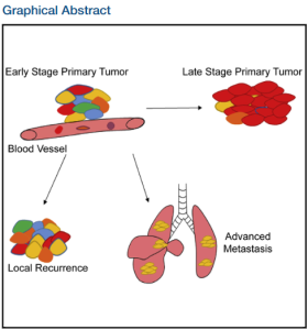
Graphical Abstract from Y.J. Tang et al., 2019 where you can see that there is a heterogenous mix of cell types in the primary tumor, but a single type in advanced metastasis.

This summer I am working to investigate what this filter might be. Using a microfluidic device, or a small device that has microchannels and chambers which cells can be seeded into, I will be studying the physical properties of MICs. One of the first things I will be considering is the process of extravasation, where MICs must travel through microbial spaces in the extracellular matrix. Using a microfluidic device with varying dimensions of microchannels, hopefully, I might be able to identify both the deformability of UPS MICs (how much they can deform to squeeze through a small gap) and if only MICs are able to deform to get through certain size channel. This may demonstrate a potential point in metastasis where the filter may lie.

Continue reading

Finally at the Starting Line

Hi everyone! I am Sid Ghanta and I’m excited to both experience and blog about my summer with BSURF.

This summer I am working with the Varghese Lab, although I have been there for some time now. There, I’ve been learning about and investigating different phenomena in the body using Organ-on-chip models, a method that creates a miniaturized 3-D cell environment mimicking tissues in the body. Moreover, it incorporates microfluidic channels, i.e. extremely tiny channels that allow fluid to move through them, that can be used to simulate movements in the body, such as blood flow or breathing. I’ve worked with numerous types of chips that seek to model areas of the body or different types of cell-cell interactions, each with unique features that aim to mimic different things, or maybe the same thing but in a new way.

I’ve always had an interest in biology and the inner workings of the body. Perhaps it sounds more philosophical than it is, but I was always curious what life was; how was it possible that everything in the body moves the way it does so perfectly and can exist in so many relatively unique forms. Additionally, as a kid, I was always quite handy with taking things apart and putting things together which is where my interest in the more mechanical side of the body was borne. This is what drew me to organ-on-chip research, a model that is so dynamic yet functional, which I hope to use in the near or distant future to investigate oncolytic virology, a long-standing interest of mine.

This summer, I am getting to focus on a specific investigation using this model and I am excited to apply what I have learned and learn some more; I finally feel like I am at the starting line of research after quite a bit of preperation. To be perfectly honest, I don’t know what to expect from this because I have never dedicated this much time to the lab, but I know I want to learn and have fun doing it. Fortunately, this summer isn’t limited to just me and the lab, so I’m expecting a great time making new friendships, making new memories, and hopefully, making new discoveries.