Our Research Tools
Duke houses a state-of-the-art Titan Krios cryo-EM microscope for data collection and a Tundra cryo-TEM for grid screening. We also have routine access to screening cryo-EM microscopes at the University of North Carolina at Chapel Hill (UNC-Chapel Hill) and National Institute of Environmental Health Sciences (NIEHS). Our lab houses a bank of FPLC systems for protein purification and construct screening (fluorescence detection), a Leica EM GP2 for cryo-EM grid preparation, and a personal server for efficient cryo-EM data processing (28 GPU cards and 600 TB storage capacity).
Protein Preparation Resources

The workhorses of the lab: Our bank of ÅKTA FPLC systems allow for up to 5 simultaneous purification runs.
Functional Characterization Resources
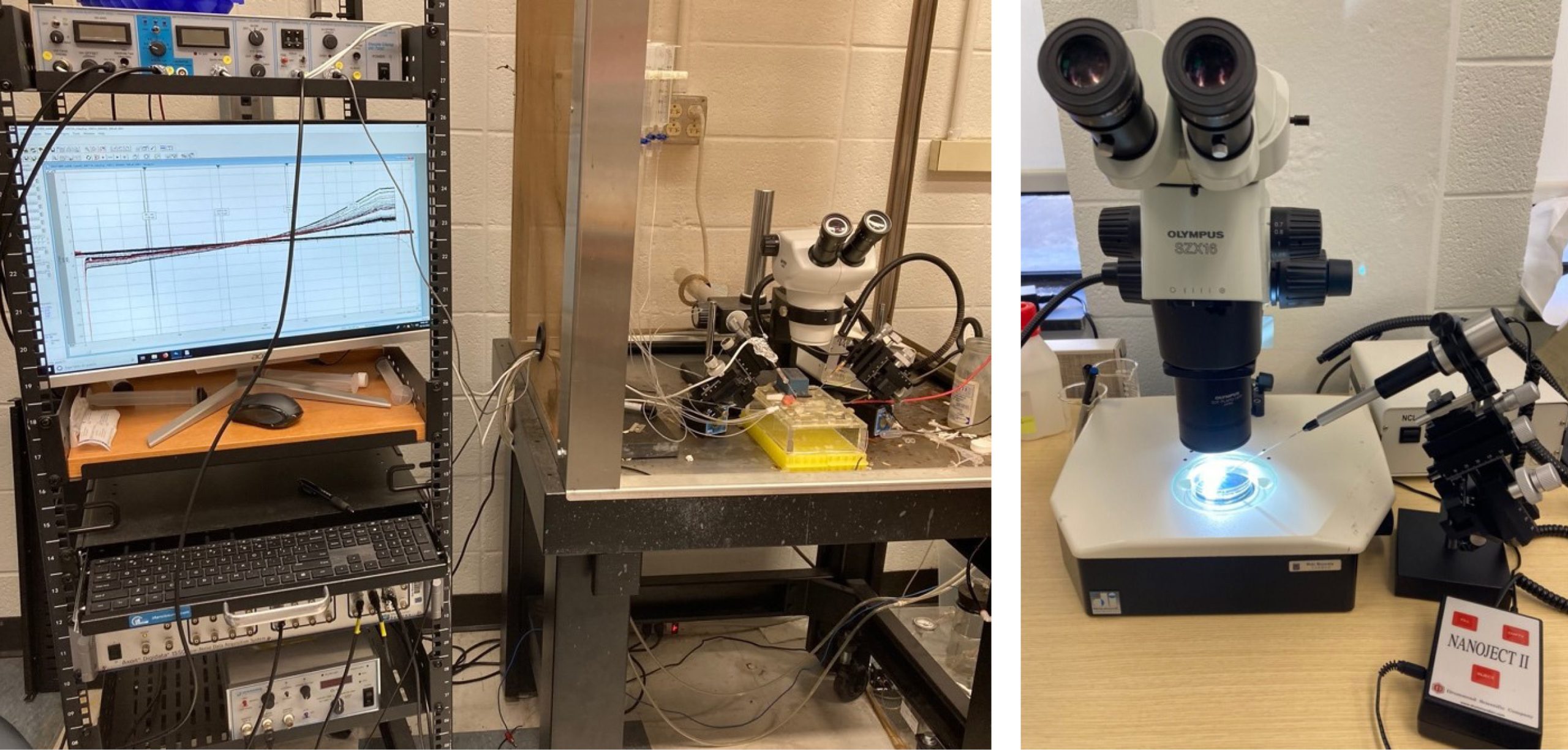
Probing ion channel and transporter function would not be possible without our two-electrode voltage clamp (TEVC) electrophysiology and oocyte injection setups.
Cryo-EM Resources

Leica EM GP2 freezing station and our own GPU server, both housed in the lab means we can go from cryo-EM grid prep to data processing all in house.
Cryo-EM Facilities
The Research Triangle Molecular Microscopy Consortium
Shared Materials Instrumentation Facility (Duke University, NC) – Thermo-Fisher Krios G3i and TUNDRA TEMs
Website: https://smif.pratt.duke.edu/node/55048
UNC – Chapel Hill CryoEM Core (University of North Carolina – Chapel Hill, NC) – Thermo-Fisher Talos Arctica TEM
Website: https://www.med.unc.edu/cryo-em/
NIEHS CryoEM Facility (NIEHS, NC) – Thermo-Fisher Talos Arctica TEM
Website: https://www.niehs.nih.gov/research/atniehs/facilities/mmc/index.cfm
External Facilities
Pacific Northwest Cryo-EM Center (NIH, OHSU & PNNL, OR)
Website: https://pncc.labworks.org/
New York Structural Biology Center (New York, NY)
Website: https://nysbc.org/semc-dept/semc-department/