
The guts of neonatal mammals and stomachless fish have a limited capacity for luminal protein digestion, which allows oral acquisition of antibodies and antigens. However, how dietary protein is absorbed during critical developmental stages when the gut is still immature remained unknown. In our study, we have found that neonatal mamamals and zebrafish have specialized intestinal cells that internalized dietary proteins and digest them intracellularly. These cells are highly endocytic and have a giant lysosomal vacuole. Thus, we named them lysosome-rich enterocytes (LREs).
We found that LREs mediate protein uptake via receptor-mediated and fluid-phase endocytosis for intracellular digestion and trans-cellular transport via a conserved endocytic machinery composed of Cubilin, Amnionless and Dab2. This machinery is required for dietary protein uptake by LREs and for growth and/or survival of larval zebrafish and mouse.
Based on our findings on the digestive functions and conserved molecular mechanisms in LREs, current projects aim to further understand cellular and molecular processes involving LREs in the context of nutrient sensing, host-microbiota interaction and immune development.

Differences in protein uptake in adult vs. neonatal mammalian intestine.

Top cartoon is depicting gavage approach for studying luminal cargo uptake into LREs (cyan box) in the larval zebrafish intestine. Lower panels are live confocal image of a 6dpf larva gavaged with fluid-phase cargo lucifer yellow (LY) and mCherry protein. Cyan box indicates magnified inset of LREs in lower panels.
Whole gut confocal image of a 6dpf Tg(ifabp:sNgly-RFP) larva secreting RFP in the anterior gut. Secreted RFP gets internalized and accumulated in LREs and then are found in other organs such as pronephros (equivalent to kidney) or liver (not shown here) over time. This suggests that LREs mediate the transport of intact proteins to other organs through transcytosis.

Electron Microscopy images of an intestinal epithelial cell (IEC) and LRE. Arrowheads mark endosomes and an arrow the lysosomal vacuole. N = nucleus, V= vacuole.

Live confocal images of LREs of a 6dpf larvae expressing GFP-Rab32a that were gavaged with mCherry. Luminal protein (mCherry) was internalized, migrated through apical endosomes (arrowheads), and progressively accumulated in LRE vacuoles (arrows).
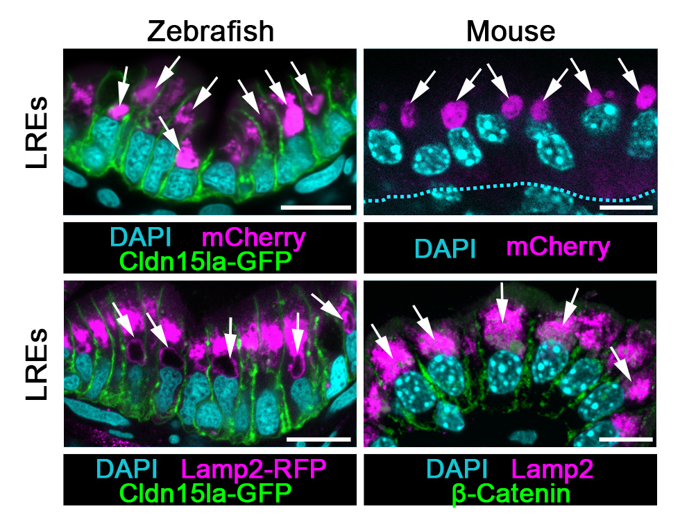
Confocal images of LREs of TgBAC(cldn15la-GFP) zebrafish larva and of P7 neonatal mouse showing that they are functionally and morphologically conserved. Arrows point to lysosomal vacuoles.