
The vertebral column or spine is a segmented structure made of alternating vertebral bodies (centra) and intervertebral discs (IVDs) that are assembled around the notochord. In our work in zebrafish we have found that the epithelial layer surrounding the notochord (the sheath) segments into alternating domains corresponding to the prospective centra and IVDs that play an instructive role in spine patterning. Current projects are aimed at understanding the cellular and molecular processes controlling notochord sheath and spine segmentation.

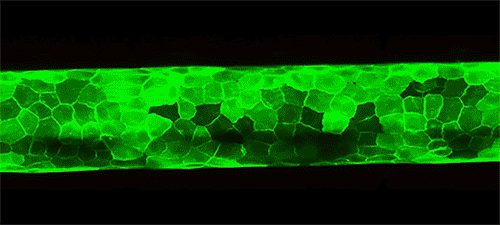
Live imaging of notochord sheath segmentation before vertebral body formation. Alternating domains are labeled with the fluorophores pkRED and GFPCaaX.
Movie shows live imaging of notochord sheath segmentation (labeled with GFPCaaX) and vertebral body formation (labeled with mCherry) over the course of spine development.

Before vertebral body formation, the notochord sheath segments into alternating domains (labeled with fluorophores pkRED and GFPCaaX). Later, osteoblasts (labeled with BFP) are specifically recruited to pkRED segments to form the vertebral bodies of the segmented spine.

When notochord segmentation is altered, parallel defects in spine segmentation arise during development. The images above show vertebral columns stained with Alizarin Red. The first and third image are spines from control animals, which display a normal pattern of spine segmentation. However, the second and fourth image are spines from animals which overexpressed Notch signaling specifically in the notochord sheath. This manipulation resulted in defects in vertebral patterning and vertebral column segmentation reminiscent of spinal defects observed in humans with Notch mutations.