
Most internal organs are networks of interconnected tubes that transport fluids. The transport of ions, water and various substances across body compartments depends on the ability of epithelial cells within tubes to develop and maintain a polarized distribution of channels, pores and transporters. On the other hand, membrane polarization is also intimately linked to the tubulogenesis process. Using zebrafish as a model system our laboratory follows an integrated approach combining forward and reverse genetics and genomics to study tube formation in the gut.
Single lumen formation:
Although tubes develop in a variety of ways, a defining characteristic of a tube is the presence of a single central lumen. We use the zebrafish gut as a model to investigate the process of lumen formation. We have previously shown that fluid accumulation is required for the enlargement and coalescence of multiple small lumens. Currently, we are investigating additional mechanisms that regulate cellular rearrangements during the process of single lumen formation.Epithelial polarization and apical membrane biogenesis:
The biogenesis of the apical surface is of particular interest in lumen formation of organs during development and defects in apical protein sorting have been linked to the etiology of numerous diseases. We are interested in determining how biogenesis of the apical surface occurs by following a comprehensive approach combining forward genetics in zebrafish and cell biological methods to elucidate genes involved in apical membrane biogenesis and lumen formation.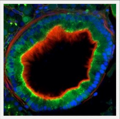

Cross section of a 4 dpf gut with apical endosomes labeled with GFP
Live confocal image of a 5 dpf zebrafish gut with an RFP-tagged apical protein and a GFP-tagged basolateral protein

An O-glycosylated protein tagged with GFP is sorted to the lumenal surface of the gut in a 5 dpf larva