Mission: Better understand the brain mechanisms in individuals experiencing post traumatic stress disorder (PTSD) and mild traumatic brain injury (mTBI) using neuroimaging tools and cognitive testing in order to identify targets for interventions or treatments.
A whole-brain voxel-based analysis of structural abnormalities in PTSD: An ENIGMA-PGC study
See CRZ, Si S, Baird CL, Haswell CC, Hussain A,…Radua J, Salminen LE, Jahanshad N, Thomopoulos SI, James A, Valmaggia L, Thompson PM, Morey RA, Kempton MJ.

In this large-scale voxel-based morphometry (VBM) meta-analysis of structural MRI data from 36 cohorts, individuals with PTSD showed significantly smaller gray matter volumes in the frontal and temporal lobes and the cerebellum, with the most pronounced reduction in the left cerebellum. White matter volume in the cerebellum was also reduced. These differences remained even when compared to trauma-exposed controls, suggesting they are specific to PTSD rather than trauma exposure alone. Along with these findings, greater PTSD severity was associated with smaller cerebellar gray matter volume, while depression severity was linked to reduced volume in both the cerebellum and superior frontal gyrus. These findings highlight the cerebellum’s emerging role in PTSD-related brain alterations and suggest that regional brain volume reductions may reflect symptom severity. Read the full article in European Psychiatry Journal.
The neurobiology of human fear generalization: A meta-analysis
Ryan D. Webler, Hannah Berg, Kimberly Fhong, Lauri Tuominen, Daphne J. Holt, Rajendra A. Morey, Iris Lange, Philip C. Burton, Miquel Angel Fullana, Joaquim Radua, Shmuel Lissek
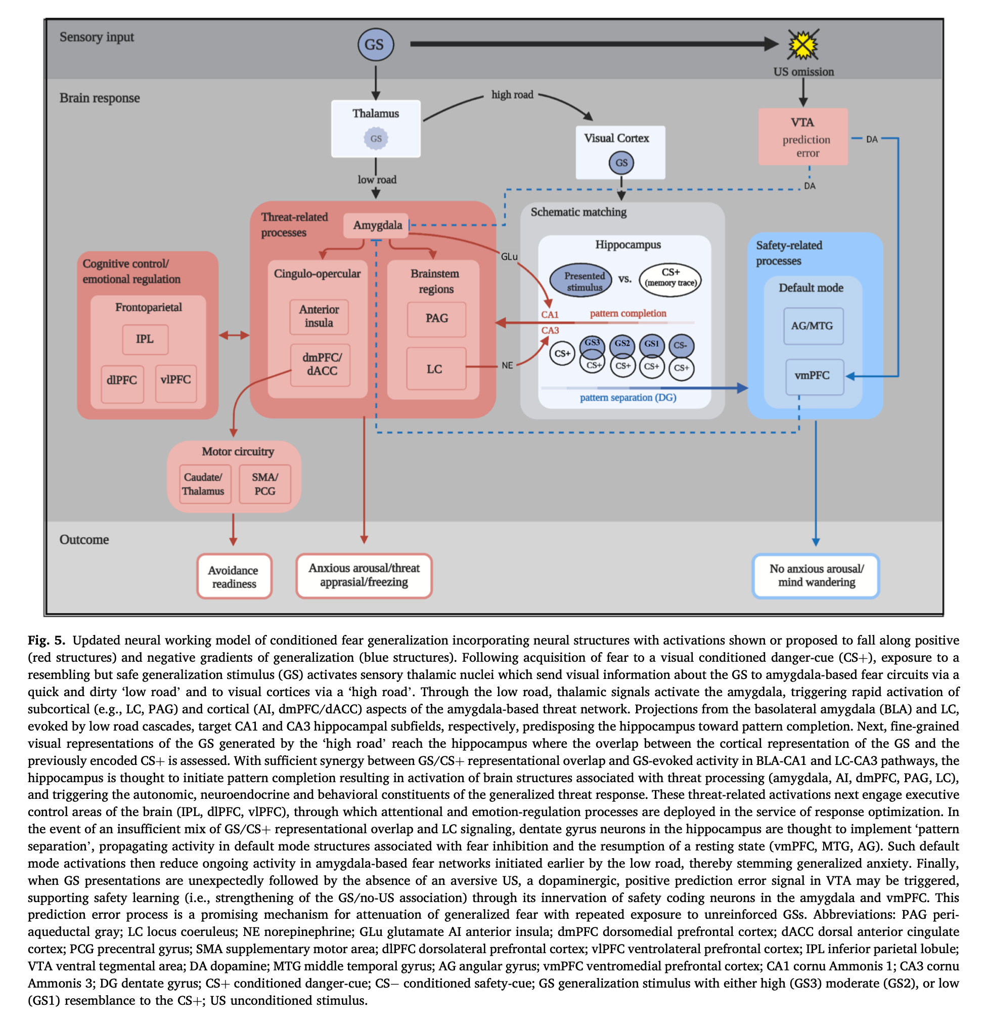
This study provides the first meta-analysis of this growing literature to delineate brain substrates of conditioned fear-generalization and formulate a working neural model. Included studies (K = 6, N = 176) reported whole-brain fMRI results and applied generalization-gradient methodology to identify brain activations that gradually strengthen (positive generalization) or weaken (negative generalization) as presented stimuli increase in CS+ resemblance. Positive generalization was instantiated in cingulo-opercular, frontoparietal, striatal-thalamic, and midbrain regions (locus coeruleus, periaqueductal grey, ventral tegmental area), while negative generalization was implemented in default-mode network nodes (ventromedial prefrontal cortex, hippocampus, middle temporal gyrus, angular gyrus) and amygdala. Findings are integrated within an updated neural account of generalization centering on the hippocampus, its modulation by locus coeruleus and basolateral amygdala, and the excitation of threat-or-safety-related loci by the hippocampus. Read the full article in Neuroscience and Biobehavioral Reviews.
FreeSurfer-based segmentation of hippocampal subfields: A review of methods and applications, with a novel quality control procedure for ENIGMA studies and other collaborative efforts
Philipp G. Sämann, Juan Eugenio Iglesias, Boris Gutman, Dominik Grotegerf, Ramona Leenings, Claas Flint, Udo Dannlowski, Emily K. Clarke-Rubright, Rajendra A. Morey, Theo G.M. van Erp, Christopher D. Whelan, Laura K. M. Han, Laura S. van Velzen, Bo Cao, Jean C. Augustinack, Paul M. Thompson, Neda Jahanshad, Lianne Schmaal
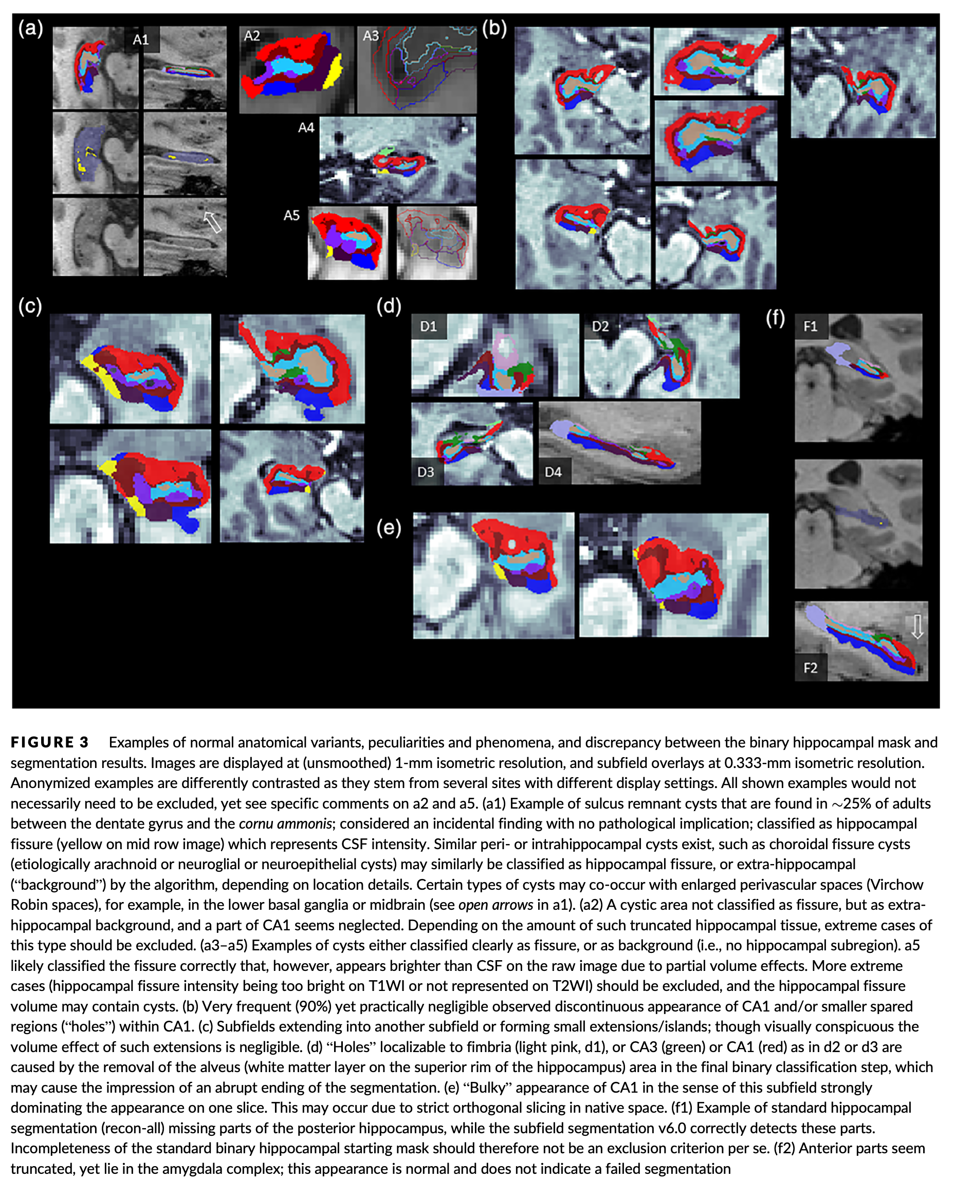
Structural hippocampal abnormalities are common in many neurological and psychiatric disorders, and variation in hippocampal measures is related to cognitive performance and other complex phenotypes such as stress sensitivity. Hippocampal subregions are increasingly studied, as automated algorithms have become available for mapping and volume quantification. In the context of the Enhancing Neuro Imaging Genetics through Meta Analysis Consortium, several Disease Working Groups are using the FreeSurfer software to analyze hippocampal subregion (subfield) volumes in patients with neurological and psychiatric conditions along with data from matched controls. In this overview, we explain the algorithm’s principles, summarize measurement reliability studies, and demonstrate two additional aspects (subfield autocorrelation and volume/ reliability correlation) with illustrative data. We then explain the rationale for a stan- dardized hippocampal subfield segmentation quality control (QC) procedure for improved pipeline harmonization. To guide researchers to make optimal use of the algorithm, we discuss how global size and age effects can be modeled, how QC steps can be incorporated and how subfields may be aggregated into composite volumes. Read the full article in Human Brain Mapping.
Volumetric trajectories of hippocampal subfields and amygdala nuclei influenced by adolescent alcohol use and lifetime trauma
Rachel D. Phillips, Michael D. De Bellis, Ty Brumback, Ashley N. Clausen, Emily K. Clarke-Rubright, Courtney C. Haswell and Rajendra A. Morey

We investigated the conditional main and interactive effects of alcohol use, and youth trauma reported at baseline, on the structural trajectories of hippocampal subfields and amygdala nuclei across adolescent development in a longitudinal sample. Greater alcohol use was associated with smaller whole hippocampus and left hippocampal tail, but larger right CA3 head and left subiculum volumes. Greater alcohol use was associated with a larger volume of the right basal nucleus of the amygdala. The effect of traumatic life events measured at the baseline visit was associated with larger right CA3 head volume in the hippocampus. Baseline trauma and within-person age change interacted such that younger adolescents with greater trauma exposure at baseline had smaller left hippocampal subfield volumes in the subiculum and molecular layer head. Read the full article in Translational Psychiatry.
Assessment of neuropsychological function in veterans with blast-related mild traumatic brain injury and subconcussive blast exposure
Ashley N. Clausen, Heather C. Bouchard, VA Mid-Atlantic MIRECC Workgroup, Kathleen A. Welsh-Bohmer and Rajendra A. Morey

We found that veterans in the combat-related blast mTBI group exhibited slower processing speed relative to controls even when controlling for PTSD and depression. Cognition did not significantly differ between subconcussive and control groups of subconcussive and combat-related blast mTBI groups. Results suggest neurocognitive assessment may not be sensitive enough to detect long-term effects of subconcussive blast exposure, or that psychiatric symptoms may better account for cognitive sequelae following combat-related subconcussive blast exposure or combat-related blast mTBI. Read the full article in Frontiers in Psychology .
Neural correlates of conceptual-level fear generalization in posttraumatic stress disorder
Rajendra A. Morey, Courtney C. Haswell, Daniel Stjepanović, VA Mid-Atlantic MIRECC Workgroup, Joseph E. Dunsmoor, Kevin S. LaBar
 We found that PTSD was associated with an enhanced neural response in fronto-limbic, midline, and occipitotemporal regions to a learned representation of threat that is based on previously established conceptual knowledge of the relationship between basic-level exemplars within a semantic category. Behaviorally, veterans with PTSD were somewhat slower to differentiate threat and safety categories as compared with trauma-exposed veteran controls owing in part to an initial overgeneralized behavioral response to the safe category. Read the full article in Neuropsychopharmacology.
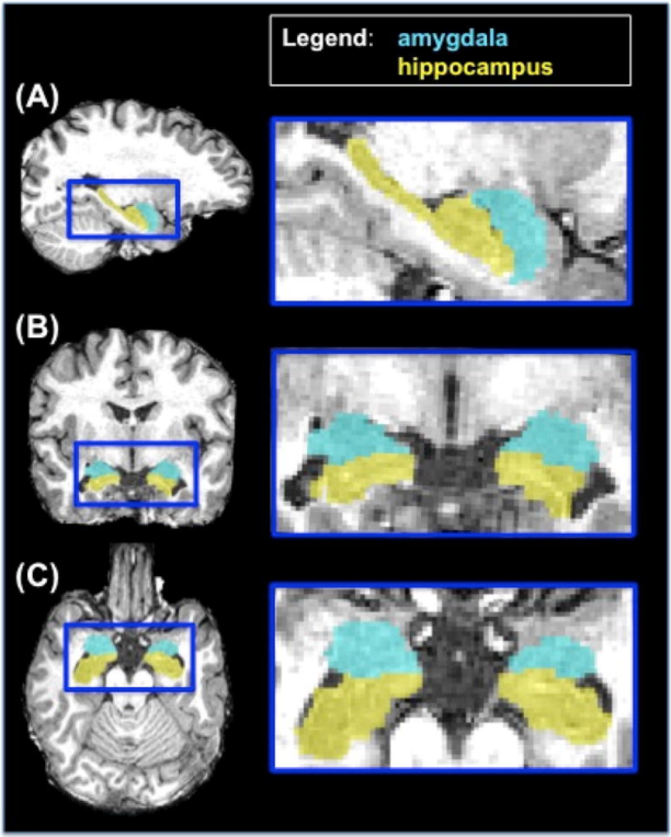
Amygdala nuclei volume and shape in military veterans with posttraumatic stress disorder
Rajendra A. Morey, Emily C. Clarke, Courtney C. Haswell, Rachel D. Phillips, Ashley N. Clausen, Mary S. Mufford, Zeynep Saygin, VA Mid-Atlantic MIRECC Workgroup, H. Ryan Wagner, Kevin S. LaBar
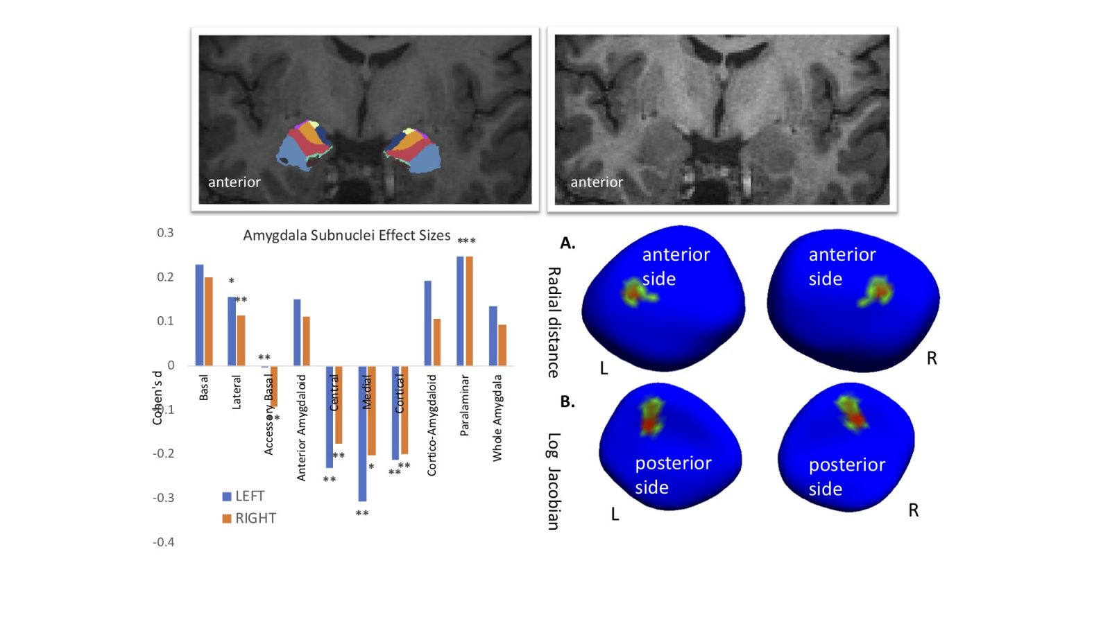 Alternations in specific amygdala subregion volumes and regional shape distortions are associated with PTSD in trauma-exposed military veterans. Volume differences of the lateral and centromedial nucleus are associated with PTSD and imply a subregion-specific pattern that is consistent with the functional roles of those two nuclei in fear learning and fear expression behaviors. Read the full article in Biological Psychiatry: Cognitive Neuroscience and Neuroimaging.
Combat exposure, posttraumatic stress disorder, and head injuries differentially relate to alterations in cortical thickness in military Veterans
Ashley N. Clausen, Emily Clarke, Rachel D. Phillips, Courtney Haswell, VA Mid-Atlantic MIRECC Workgroup, & Rajendra A. Morey
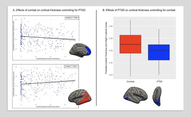
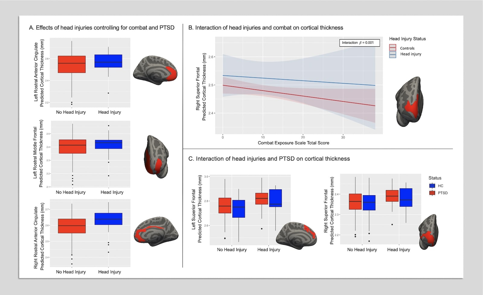
These results may indicate that combat exposure severity, PTSD, and prior head injuries have differential impacts on cortical thickness in Veteran populations. Utilization of longitudinal research designs and implementation of multimodal neuroimaging approaches (i.e., functional and structural assessments) are warranted and will clarify the functional implications of the present findings. Read the full article in Neuropsychopharmacology.
Resting-state brain fluctuation and functional connectivity dissociate moral injury from posttraumatic stress disorder
Delin Sun , Rachel D. Phillips, Hannah L. Mulready, Stephen T. Zablonski, Jessica A. Turner, Matthew D. Turner, Kathryn McClymond, Jason A. Nieuwsma, and Rajendra A. Morey.
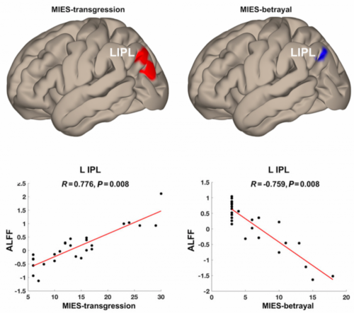
Moral injury is closely associated with posttraumatic stress disorder (PTSD) and characterized by disturbances in social and moral cognition. Our results provide the first evidence that morally injurious events and PTSD symptoms have dissociable neural underpinnings, and that behaviorally distinct subcomponents of morally injurious events are different in neural responses. Read the full article in Depression & Anxiety.
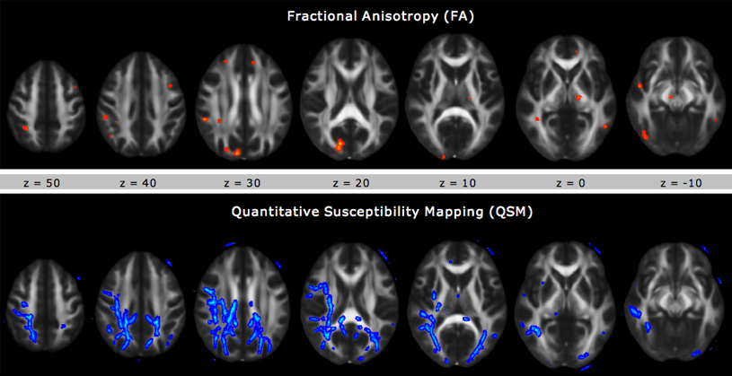
Due to its ability to identify myelin rich areas in the brain, Quantitative Susceptibility Mapping (QSM) provides the ability to isolate specific components of white matter damage and gives a better idea of myelin integrity than Diffusion Tensor Imaging (DTI) or Fractional Anisotropy (FA). As seen in the figure, QSM imaging reveals more TBI associated differences than FA.
Brain structural covariance network centrality in maltreated youth with PTSD and in maltreated youth resilient to PTSD
Delin Sun , Courtney C. Haswell, Rajendra A. Morey, and Michael D. De Bellis
 Child maltreatment is a major cause of pediatric posttraumatic stress disorder (PTSD). This work is the first to identify cortical thickness-based structural covariance network differences between maltreated youth with and without PTSD. Network differences are demonstrated in both networks unique to maltreated youth with PTSD and those resilient to PTSD. The networks identified are important for the successful attainment of age-appropriate social cognition, attention, emotional processing, and inhibitory control.
Fear learning circuitry is biased toward generalization of fear associations in posttraumatic stress disorder
RA Morey, JE Dunsmoor, CC Haswell, VM Brown, A Vora, J Weiner, D Stjepanovic, HR Wagner III, VA Mid-Atlantic MIRECC Workgroup, and KS LaBar

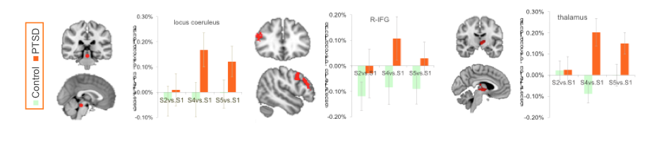
Compared to trauma-exposed controls, PTSD patients exhibited more severe memory distortion of the fear-conditioned stimulus biased toward the face-stimulus expressing the highest fear intensity. PTSD patients exhibited a neural activation bias – greater activation that generalized toward the face-stimuli expressing higher fear intensity than the conditioned stimulus. Differential activation can be seen in various brain regions, including the locus coeruleus, inferior frontal gyrus, and thalamus. Read the article in Translational Psychiatry.
Amygdala volume changes with posttraumatic stress disorder in a large case-controlled veteran group
Rajendra A. Morey, M.D., M.S., Andrea L. Gold, M.S., Kevin S. LaBar, Ph.D., Shannon K. Beall, B.S., Vanessa M. Brown, B.A., Courtney C. Haswell, M.S., Jessica D. Nasser, Mid-Atlantic MIRECC Workgroup, H. Ryan Wagner, Ph.D., and Gregory McCarthy, Ph.D.
PTSD is associated with atrophy of the hippocampus and amygdala in a large group (n = 186) of recent veterans from Iraq and Afghanistan. Furthermore, the duration of illness appears to modulate the extent of atrophy. Read the article in Archives of General Psychiatry.
Neural systems for guilt from actions affecting self versus others
Rajendra A. Morey, Gregory McCarthy, Elizabeth S. Selgrade, Srishti Seth, Jessica D. Nassera, and Kevin S. LaBar
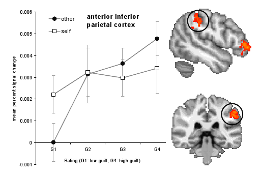 Specific brain regions are more highly activated for feelings of guilt when an individuals actions lead to harming others compared to harming only oneself. Regions such as the anterior inferior parietal (AIP) cortex, the anterior inferior frontal gyrus (IFG), and the ventromedial prefrontal cortex are differentially involved. Read the article in NeuroImage.
Staying cool when things get hot: emotion regulation modulates neural mechanisms of memory encoding
Jasmeet Pannu Hayes, Rajendra A. Morey, Christopher M. Petty, Srishti Seth, Moria J. Smoski, Gregory McCarthy, and Kevin S. LaBar
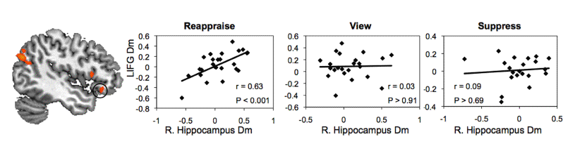 During times of emotional stress, individuals often engage in emotion regulation to reduce the experiential and physiological impact of negative emotions. Neurobehavioral evidence shows that engaging in cognitive reappraisal is advantageous to both affective and mnemonic processes. Read the article in Frontiers.
Effects of chronic mild traumatic brain injury on white matter integrity in Iraq and Afghanistan war veterans
Rajendra A. Morey, Courtney C. Haswell, Elizabeth S. Selgrade, Dino Massoglia, Chunlei Liu, Jonathan Weiner, Christine E. Marx, MIRECC Work Group, Ibolja Cernak, and Gregory McCarthy
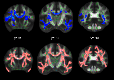
Our results with DTI whole brain crossing fiber analysis shows that the duration of loss of consciousness (Blue Areas) and feeling dazed and confused (Pink Areas) predicts disruption of white matter in mild TBI. Read the article in Human Brain Mapping.
A comparison of automated segmentation and manual tracing for quantifying hippocampal and amygdala volumes
Rajendra A. Morey, Christopher M. Petty, Yuan Xu, Jasmeet Pannu Hayes, H. Ryan Wagner II, Darrell V. Lewis, Kevin S. LaBar, Martin Styner, Gregory McCarthy
 Shape analysis using surface mesh obtained with spherical harmonics, shows systematic differences between two popular segmentation tools (FreeSurfer and FIRST) and gold standard hand tracing of the hippocampus particularly in the head and tail sections. Read the article in NeuroImage.
Intermittent Theta Burst Stimulation (iTBS) and Functional Connectivity in People Living with HIV/AIDS (PLWHA) Who Smoke Tobacco Cigarettes: A Preliminary Pilot Study. Accepted.
Rakesh G, Adams TG, Morey RA, Alcorn III JL, Khanal R, Su AE, Himelhoch SS and Rush CR.