A Virtual Symposium hosted by the Fitzpatrick Institute for Photonics | March 7-8, 2022
Poster Session
There are virtual recorded presentations by some of the designated posters.
Please look for the BLUE STAR for those have recorded their poster presentation.
Click a poster image thumbnail to enlarge.
1. Synergistic Immuno Photothermal Nanotherapy (SYMPHONY) for Effective Cancer Treatment
Virtual Poster Presentation: Yang Liu


Yang Liu, Brant A. Inman, Gregory M. Palmer, Peter E. Fecci, and Tuan Vo-Dinh
Department of Biomedical Engineering, Duke University
Activating the immune system has long been a goal in oncology, and recently immunotherapy has emerged as one of the most promising cancer treatment breakthroughs. We have developed an innovative cancer therapy named as Synergistic Immuno Photothermal Nanotherapy (SYMPHONY) by combining gold nanostars (GNS)-mediated photothermal ablation with checkpoint inhibitor immunotherapy. GNS nanoparticle has multiple sharp branches for tip-enhanced plasmonics and tunable plasmonic absorption in the near-infrared (NIR) tissue optical window. GNS has superior photon-to-heat conversion capability for effective photothermal therapy with NIR laser. Our group has innovated a toxic chemical-free method to synthesize GNS nanoparticles and apply the biocompatible GNS nanoparticles for SYMPHONY therapy. In vivo experiment with brain cancer and bladder cancer murine animal models demonstrate that our superior GNS-mediated photothermal therapy dramatically amplifies the anti-cancer immune response in synergy with checkpoint blockade immunotherapy. SYMPHONY therapy results in not only primary tumor shrinkage but also recurrence prevention, implying the generation of an anti-cancer vaccine effect. As a result, our novel SYMPHONY therapy has the potential to substantially improve outcomes of brain cancer patients in future clinical applications.
yang.liu3@duke.edu
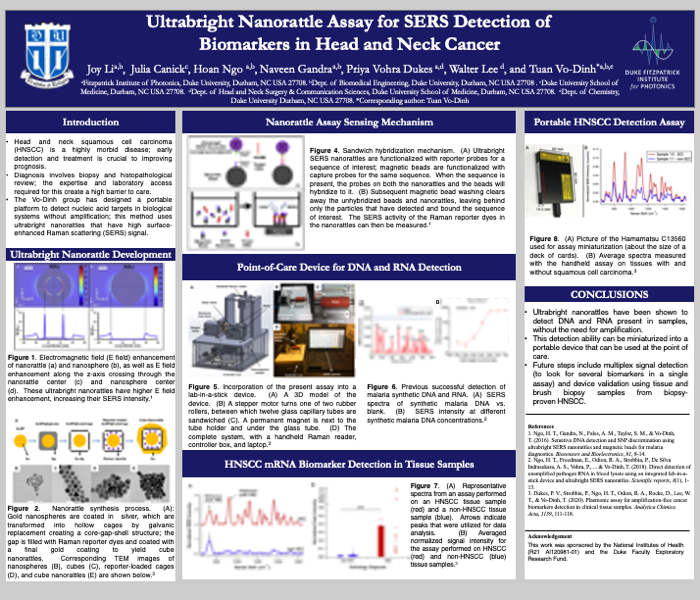
2. Ultrabright Nanorattle Assay for SERS Detection of Biomarkers in Head and Neck Cancer
Joy Li, Julia Canick, Hoan Ngo, Naveen Gandra, Priya Vohra Dukes, Walter Lee, and Tuan Vo-Dinh
Fitzpatrick Institute of Photonics, Duke University, Durham, NC USA 27708.
Dept. of Biomedical Engineering, Duke University, Durham, NC USA 27708 .
Duke University School of Medicine, Durham, NC USA 27708.
Dept. of Head and Neck Surgery & Communication Sciences, Duke University School of Medicine, Durham, NC USA 27708.
Dept. of Chemistry, Duke University Durham, NC USA 27708.
Head and neck squamous cell carcinoma (HNSCC) is the sixth most common cancer worldwide; though it has a high morbidity and mortality, rapid diagnosis and treatment can often be curative. In low-to-middle-income countries, however, this process is often delayed due to inadequate resources and personnel. A promising route of diagnosis is the detection of HNSCC-associated mRNA biomarkers. Surface-enhanced Raman spectroscopy (SERS) is effective for the recognition of nucleic acid disease markers without amplification and provides a potential avenue for investigation of rapid diagnostic methods. Our group has developed ultrabright “nanorattles,” with a structural gap loaded with Raman reporter dyes. These nanorattles, loaded with reporter probes, work in concert with magnetic beads, loaded with capture probes. When the sequence of interest is detected, the nanorattles and the magnetic beads hybridize to the mRNA; after the unbound particles are washed away, the hybridized sandwich assay is concentrated on a spot and its SERS signal is measured. This method has previously been successful in detecting malaria RNA and synthetic DNA. We also found that the assay can detect HNSCC in tissue samples from biopsy-proven head and neck squamous cell carcinoma; this detection has high diagnostic accuracy, with a sensitivity of 100% and specificity of 97%. SERS-based assays are a promising option for HNSCC diagnostics. A rapid diagnostic device employing SERS methods has the potential to expedite the timely diagnosis and treatment of head and neck squamous cell carcinomas in LMICs.
jql5@duke.edu
3. Intracranial accumulation of plasmonic gold nanostars for the acceleration of tumor laser ablation

Ethan S. Srinivasan, Pakawat Chongsathidkiet, Ren A. Odion, Yang Liu, Eric W. Sankey, Ryan M. Edwards, Tuan Vo-Dinh, Peter E. Fecci
School of Medicine, Duke University
Background: Laser interstitial thermal therapy (LITT) is an effective minimally-invasive treatment option for intracranial tumors. Our group produced plasmonics-active gold nanostars (GNS) designed to preferentially accumulate within intracranial tumors and amplify the ablative capacity of LITT while protecting surrounding tissue. Methods: The 12nm GNS were synthesized using reduced HAuCL4 with Na3C6H5O7 seeds, mixed with AgNO3, C6H8O6, and HAuCL4, and coated with polyethylene glycol then functionalized with methoxy PEG thiol. CT-2A glioma cells were intracranially implanted into mice, followed 18 days later by IV injection of GNS. PET-CT was performed at 10-minutes, 24-, and 72-hours post-GNS administration, with autoradiography (AR) and histopathology (HP) on sacrifice after the last scan. To test the impact of GNS on LITT coverage capacity in appropriately sized ex vivo models, we utilized agarose gel-based phantoms incorporating control and GNS-infused central “tumors” in multiple shapes. LITT was administered with the NeuroBlate System. Results: In vivo, GNS preferentially accumulated within intracranial tumors on PET-CT at the 24- and 72-hour timepoints. AR and HP confirmed high GNS accumulation within tumor. Ex vivo, in cuboid tumor phantoms, the GNS-infused phantom heated 5.5x faster than the control, rising 0.49°C per minute compared to 0.09°C. In a split-cylinder tumor phantom with half containing GNS, the GNS-infused border heated 2x faster and the surrounding area was exposed to 30% lower temperatures. Conclusions: Our results provide evidence for use of GNS to improve the efficiency and potentially safety of LITT. The in vivo data support selective accumulation within intracranial tumors, and the GNS-infused phantom experiments demonstrate increased rates of heating within the tumor model, heat contouring to tumor borders, and decreased heating of surrounding regions representing normal structures.
Ethan.srinivasan@duke.edu
4. Metallo-graphene enhanced upconversion luminescence for broadband photodetection under polychromatic illumination
Virtual Poster Presentation: Akash Gupta


Akash Gupta, Yong Il Park, Surojit Chattopadhyay
Institute of Biophotonics, National Yang-Ming Chiao Tung University, Taipei 112, Taiwan;
School of Chemical Engineering, Chonnam National University, Gwangju, South Korea
Here, we used electrostatically conjugated SiO2-coated upconversion nanoparticles (UCNPs), and gold nanorods (AuNRs) nanocomposite (NC) on graphene to obtain >200-fold upconversion luminescence (UCL) enhancement. Plasmonic AuNR and graphene imparted maximum plasmonic enhancement of the UCL in the UCNP with an optimized 7 nm thick SiO2 shell. This is attributed to the enhanced absorption in the UCNPs by the nano-antennae effect of the AuNRs as shown by finite difference time domain (FDTD) simulation. The enhanced UCL was directly confirmed by confocal fluorescence imaging. Finally, a NC/graphene hybrid photodetector (PD) was fabricated that showed broadband (455-980 nm) photoresponse, with photoresponsivity of ~ 5000 AW-1, and fast response times of 80 ms, compared to 3 s obtained for a device without the AuNRs. The conventional multiphoton infrared (~ 980 nm) absorbing UCNPs demonstrated an interesting high energy (blue (B), green (G), and red (R)) photoresponse that is now attributed to weak single-photon absorption in the UCNPs. This allowed us to study the performance of the hybrid PD under polychromatic illumination using individual B, G, R, and a combination of B+G, B+R, G+R, and B+G+R. The results of polychromatic illumination indicated absorption saturation in UCNPs under one-photon absorption. The device has been used for detecting signals from domestic appliances, such as frequency modulated AC remote controllers, and attributed the speed to the fast charge sweeping by the AuNRs.
gupta.akash00@gmail.com
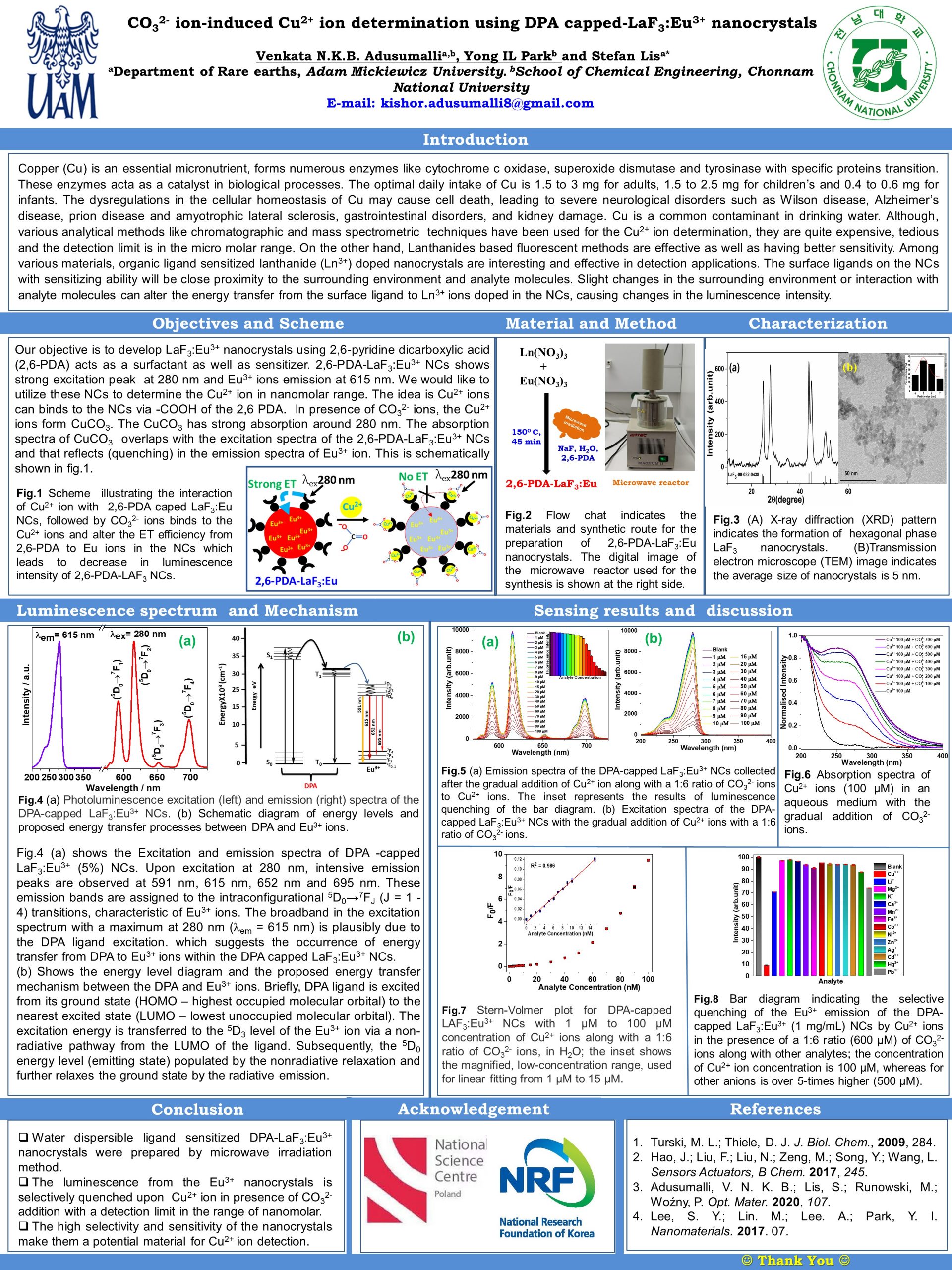
5. CO32- ion-induced Cu2+ ion determination using DPA capped-LaF3:Eu3+ nanocrystals
Virtual Poster Presentation: Venkata Adusumalli

Venkata Nanda Kishor Babu Adusumalli, Yong IL Park, Stefan Lis
(a)Department of Rare Earths, Adam Mickiewicz University, Poland
(b) School of Chemical Engineering, Chonnam National University, South Korea
In recent years, organic ligand sensitized Ln-doped nanoparticles are gained importance in sensing and determination of biomolecules, metal ions and organic and inorganic pollutants. Here we report the carbonate (CO32-) ion induced Cu2+ ion determination in aqueous medium using 2,6-pyridinedicaboxylic acid (DPA)-capped LaF3:Eu3+ nanocrystals (NCs). Copper (Cu) is an essential micronutrient, forms numerous enzymes like cytochrome c oxidase, superoxide dismutase and tyrosinase with specific proteins transition. These enzymes acta as a catalyst in biological processes. The optimal daily intake of Cu is 1.5 to 3 mg for adults, 1.5 to 2.5 mg for children’s and 0.4 to 0.6 mg for infants. The dysregulations in the cellular homeostasis of Cu may cause cell death, leading to severe neurological disorders such as Wilson disease, Alzheimer’s disease, prion disease and amyotrophic lateral sclerosis, gastrointestinal disorders, and kidney damage. Cu is a common contaminant in drinking water. In DPA-LaF3:Eu3+ NCs, DPA acts as surfactant to LaF3:Eu3+ NCs and transfer energy the Eu3+ ions via sensitization. These NCs exhibit strong red emission at 615 nm from Eu3+ ions via DPA at 280 nm excitation. In the aqueous medium the Cu2+ ions bind to LaF3 NCs via -COOH of the DPA. In presence of CO32- ions, the Cu2+ ions form CuCO3. The CuCO3 has strong absorption around 280 nm. The absorption spectra of CuCO3 overlaps with the excitation spectra of the DPA-LaF3:Eu3+ NCs and that reflects (quenching) in the emission spectra of Eu3+ ion. The quenching was highly selective only towards Cu2+ ions and devoid of any interference from several other analytes. The calculated detection limit was 117 nM.
kishor.adusumalli8@gmail.com
6. The use of porphyrins and their derivatives in photodynamic action on Staphylococci
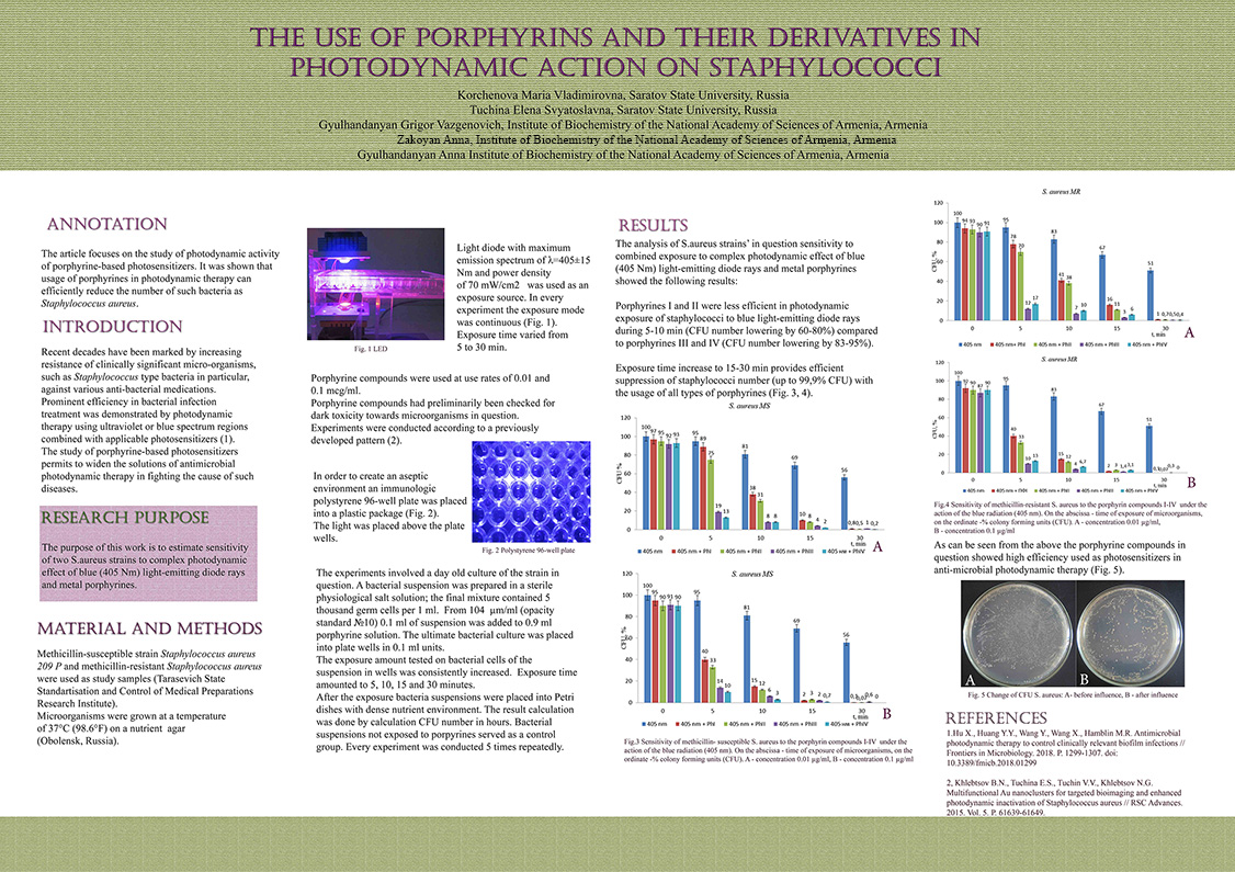
Maria V. Korchenova, Elena S. Tuchina, Dr. Grigor V. Gyulkhandanyan, Anna A. Zakoyan, Anna G. Gyulkhandanyan
Department of Biology, Saratov State University, Russia
The aim of this work was to determine and test the most effective cationic porphyrins and metalloporphyrins with high photoactivity against methicillin-resistant and methicillin-sensitive Staphylococcus aureus without attracting additional compounds that increase their effectiveness. It was shown that the synthesized cationic porphyrins/metalloporphyrins exhibit a high degree of phototoxicity towards MSSA and MRSA strains.
tuchinaes@gmail.com
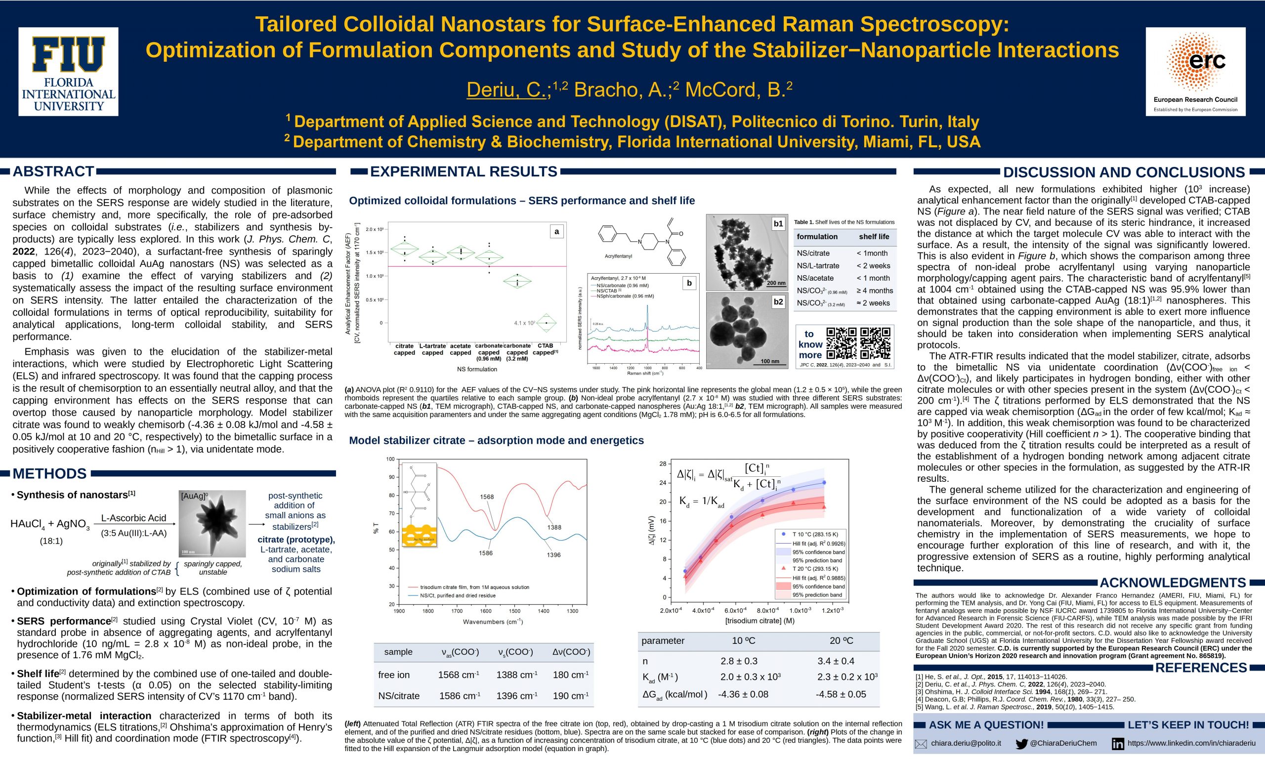
7. Tailored Colloidal Nanostars for Surface Enhanced Raman Spectroscopy (SERS): Optimization of Formulation Components and Study of the Stabilizer-Nanoparticle Interactions
Deriu, C.;(1,2) Bracho, A.;(2) McCord, B.(2)
(1) Department of Applied Science and Technology (DISAT), Politecnico di Torino. Turin, Italy;
(2) Department of Chemistry & Biochemistry, Florida International University, Miami, FL, USA
While the effects of morphology and composition of plasmonic substrates on the SERS response are widely studied in the literature, surface chemistry and, more specifically, the role of pre-adsorbed species on colloidal substrates (i.e., stabilizers and synthesis by-products) are typically less explored. In this work (J. Phys. Chem. C, 2022, 126(4), 2023−2040), a surfactant-free synthesis of sparingly capped bimetallic colloidal AuAg nanostars was selected as a basis to (1) examine the effect of varying stabilizers and (2) systematically assess the impact of the resulting surface environment on SERS intensity. The latter entailed the characterization of the colloidal formulations in terms of optical reproducibility, suitability for analytical applications, long-term colloidal stability, and SERS performance. Emphasis was given to the elucidation of the stabilizer-metal interactions, which were studied by Electrophoretic Light Scattering (ELS) and infrared spectroscopy. It was found that the capping process is the result of chemisorption to an essentially neutral alloy, and that the capping environment has effects on the SERS response that can overtop those caused by nanoparticle morphology. Model stabilizer citrate was found to weakly chemisorb (-4.36 ± 0.08 kJ/mol and -4.58 ± 0.05 kJ/mol at 10 and 20 °C, respectively) to the bimetallic surface in a positively cooperative fashion (nHill > 1), via unidentate mode.
chiara.deriu@polito.it
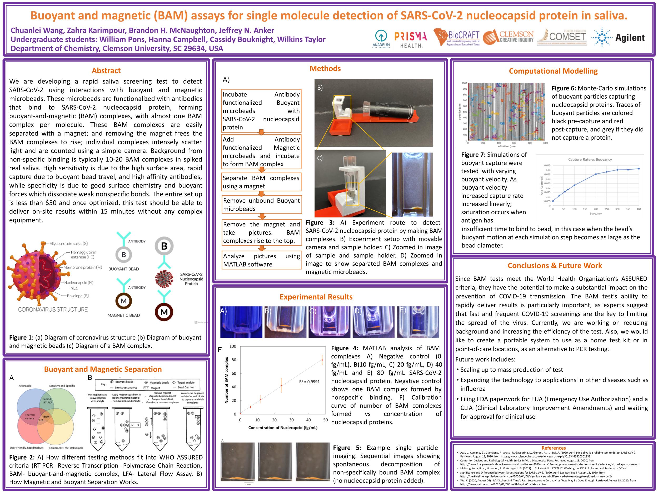
8. Buoyant and magnetic (BAM) assays for single molecule detection of SARS-CoV-2 nucleocapsid protein in saliva.
Chuanlei Wang, Will Pons*, Hanna Campbell*, Cassidy Bouknight*, Wilkins Taylor*, Zahra Karimpour, Brandon H. McNaughton, Jeffrey N. Anker *These authors contributed equally
Department of Chemistry, Clemson University
We are developing a rapid saliva screening test to detect SARS-CoV-2 using interactions with buoyant and magnetic microbeads. These microbeads are functionalized with antibodies that bind to SARS-CoV-2 nucleocapsid protein, forming buoyant-and-magnetic (BAM) complexes, with almost one BAM complex per molecule. These BAM complexes are easily separated with a magnet; and removing the magnet frees the BAM complexes to rise; individual complexes intensely scatter light and are counted using a simple camera. Background from non-specific binding is typically 10-20 BAM complexes in spiked real saliva. High sensitivity is due to the high surface area, rapid capture due to buoyant bead travel, and high affinity antibodies, while specificity is due to good surface chemistry and buoyant forces which dissociate weak nonspecific bonds. The entire set up is less than $50 and once optimized, this test should be able to deliver on-site results within 15 minutes without any complex equipment.
janker@clemson.edu
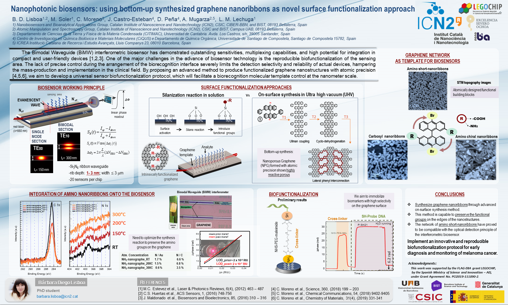
9. Nanophotonic biosensors: using bottom-up synthesized graphene nanoribbons as novel surface functionalization approach
B. D. Lisboa (1,2), M. Soler (1), C. Moreno (3), J. Castro-Esteban (4), D. Peña (4), A. Mugarza (4,5), L. M. Lechuga (1)
1) Nanobiosensors and Bioanalytical Applications Group, Catalan Institute of Nanoscience and Nanotechnology (ICN2), CSIC, CIBER-BBN, and BIST, 08193 Bellaterra, Spain;
2) Atomic Manipulation and Spectroscopy Group, Catalan Institute of Nanoscience and Nanotechnology (ICN2), CSIC and BIST, Campus UAB, 08193 Bellaterra, Spain;
3) Departamento de Ciencias de la Tierra y Fisica de la Materia Condensada (CITIMAC), Universidad de Cantabria. Avda. Los Castros, s/n, 39005 Santander, Spain;
4) Centro de Investigación en Química Biolóxica e Materiais Moleculares (CiQUS) e Departamento de Química Orgánica, Universidade de Santiago de Compostela, Santiago de Compostela 15782, Spain;
5) ICREA Institució Catalana de Recerca i Estudis Avançats, Lluis Companys 23, 08010 Barcelona, Spain.
Biosensors are profiled as next-generation diagnostics devices, offering point-of-care rapid testing with excellent performance. Among them, the Bimodal Waveguide (BiMW) interferometric biosensor has demonstrated outstanding sensitivities, multiplexing capabilities, and high potential for integration in compact and user-friendly devices. One of the major challenges in the advance of biosensor technology is the reproducible biofunctionalization of the sensing area. The lack of precise control during the arrangement of the biorecognition interface severely limits the detection selectivity and reliability of actual devices, hampering the mass-production and implementation in the clinical field. By proposing an advanced method to produce functionalized graphene nanostructures with atomic precision, we aim to develop a universal sensor biofunctionalization protocol, which will facilitate a biorecognition molecular template control at the nanometer scale.
Here, we combine the possibility to functionalize graphene building blocks with atomic precision via a bottom-up synthesis approach to support the production of different graphene nanoarchitectures (i.e., graphene nanoribbons and nanoporous graphene) containing selective anchoring groups such as amine, carboxyl and epoxy groups. The quality of the functionalized graphene template is dictated by the on-surface synthesis approach carried out in UHV (ultra-high vacuum) conditions. Additionally, the graphene successful integration on the BiMW biosensor is obtained by a direct transfer to preserve the stability of the graphene under flow conditions. Finally, as a proof of concept, nucleic acids biomarkers will be detected applying a universal biofunctionalization protocol for the early, non-invasive diagnostic of melanoma cancer.
barbara.lisboa@icn2.cat
10. Magnetic metal-inorganic composite as new multimodal contrast agents: preliminary research design

Olimpia Tammaro1, Michele Pansini2, Serena Esposito1, Laura Fabris1
1Department of Applied Science and Technology, Politecnico, Politecnico di Torino, Corso Duca degli Abruzzi 24, 10129 Torino, Italy
2Department of Civil and Mechanical Engineering, Università degli Studi di Cassino e del Lazio Meridionale, via G. Di Biasio 43, 03043 Cassino, FR, Italy
The design of new multimodal contrast agents (MCAs) is still challenging. Despite the huge interest in this research field, major progress is still needed to achieve a real and scalable process. The ideal process should be able to combine different phases within the same diagnostic tool, overcoming the additional synthetic steps and high-cost procedures. In detail, in the case of the multimodal approach to combine SERS and MRI signals, a great interest is focused on the synthesis of Au-Fe nanoalloys as MCAs, exploiting their magneto-plasmonic behaviour. However, the production of a Au-Fe nanoalloy as single unit is tricky because a precise control of particle size, composition, and surface property is required. One possible strategy is to use supports, such as zeolites, in the synthetic design to simplify the process. In this view, our idea is to design a novel strategy to obtain nanoalloys by exploiting prior knowledge in the synthesis of metal-inorganic nanocomposites. As the first step, we started studying the production of Fe NPs supported by an inorganic matrix obtained from zeolite precursor. The employed raw materials are commercial and/or natural zeolites (price per kg, some dozens of dollars). The zeolites are treated following a patented process. In detail, they are repeatedly exchanged with transition metal ions (here, Fe2+) and then subjected to chemical reduction by thermal treatment at a relatively mild temperature under reducing atmosphere. Already after the cation exchange steps, it is visible the formation of small Fe NPs embedded in the zeolite structure, while the final product, after the thermal treatment is represented by a metal-inorganic nanocomposite, where nanoparticles of Fe0/ Fe3O4 are dispersed within a ceramic matrix due to zeolite collapsed structure. The future steps involve the addition of the second metallic component (Au) during the cation exchange steps, in order to obtain an Au-Fe alloy. Once obtained this alloy, we will conduct a study on the effect of the thermal treatment to define the best applicability conditions (presence or not of an amorphous matrix). The simplicity of the process together with the versatility of the materials obtained has already produced excellent results in various fields.
olimpia.tammaro@polito.it
11. In vivo SERS monitoring in plants using plasmonic nanoprobes
Virtual Poster Presentation: Vanessa Cupil-Garcia


Vanessa Cupil-Garcia, Joy Li, Ren Odion, Dr. Pietro Strobbia, Dr. Bridget M. Crawford, Dr. Hsin-neng Wang, Dr. Jianhong Hu, Dr. Rodolfo Zentella, Dr. Kenneth Kemner, Dr. Tai-Ping Sun, and Dr. Tuan Vo-Dinh
Department of Chemistry, Duke University
Further understanding of biomass producing associated metabolic pathways in plants can be used to increase the production of biomass. In vivo detection of these markers has proved to be limited due to complex sample preparation required by traditional methods. Recently the Vo-Dinh group has designed a platform to detect nucleic acid targets in biological systems called inverse molecular sentinels (iMS) which utilize surface-enhanced Raman scattering. These multimodal probes were shown to detect and image key microRNA within whole plants in vivo.1 This work lays the foundation for detecting and imaging biological markers in plants with enhanced spatial and temporal resolution.
vanessa.cupil.garcia@duke.edu
12. Detection of Plant miRNA using Plasmonic Biosensors with Shifted Excitation Raman Difference Spectroscopy

Ren A. Odion, Pietro Strobbia, Bridget Crawford, Rodolfo Zentella, Martin Maiwald, Bernd Sumpf, Tai-ping Sun, Tuan Vo-Dinh
Ren A. Odion, Pietro Strobbia, Bridget Crawford, Rodolfo Zentella, Martin Maiwald, Bernd Sumpf, Tai-ping Sun, Tuan Vo-Dinh
The detection of micro-RNAs (miRNAs) is crucial in understanding the developmental process of key genes involved in the biomass production of plant biofuels. Current methods for understanding these pathways rely on slow methods such as polymerase chain reaction (PCR) to amplify a tediously purified sample of miRNA from plants. To this end, we have developed a combined plasmonic biosensing method based on a Surface Enhanced Raman Spectroscopy (SERS) platform called the inverse Molecular Sentinel (iMS) to directly detect miRNA such as miR858a to understand ligin production and increased biomass. This biosensor is then coupled with the Shifted Excitation Raman Difference Spectroscopy (SERDS) technique to detect these targets in the field, even in the presence of harsh background illumination. The application of such technology for monitoring plant gene expression in the field may potentially revolutionize agriculture technology through the use of nanotechnology-based monitoring for plant health, pollution, and pathogen detection.
ren.odion@duke.edu
13. Plasmonic Gold Nanostars: Synthesis and Application

Supriya Atta, Tuan Vo-Dinh
Department of Biomedical Engineering, Duke University
Among the various types of gold nanoparticle systems, gold nanostars (GNS) have been widely recognized for their ability to create strong Localized Surface Plasmon Resonances (LSPRs), which is strongly depended on the size, shape, length, and number of the spikes of the nanostars. Unfortunately, traditional protocols for gold nanostars syntheses have failed to tune the size, shape, number and length of the spikes. Moreover, there is a lack of sufficient monodispersity and reproducibility of the traditional protocols. We will discuss here a facile seed-mediated synthesis of multibranched stars to control the spike length and spike number of the nanostars. This nanostars synthesis method produces a number of different spike length, and number which can be tuned by changing the on the concentrations of the seeds, AgNO3, ascorbic acid, and surfactant. The development of gold nanostars will lead to a wide variety of applications for in vitro and in vivo biomedical diagnostics.
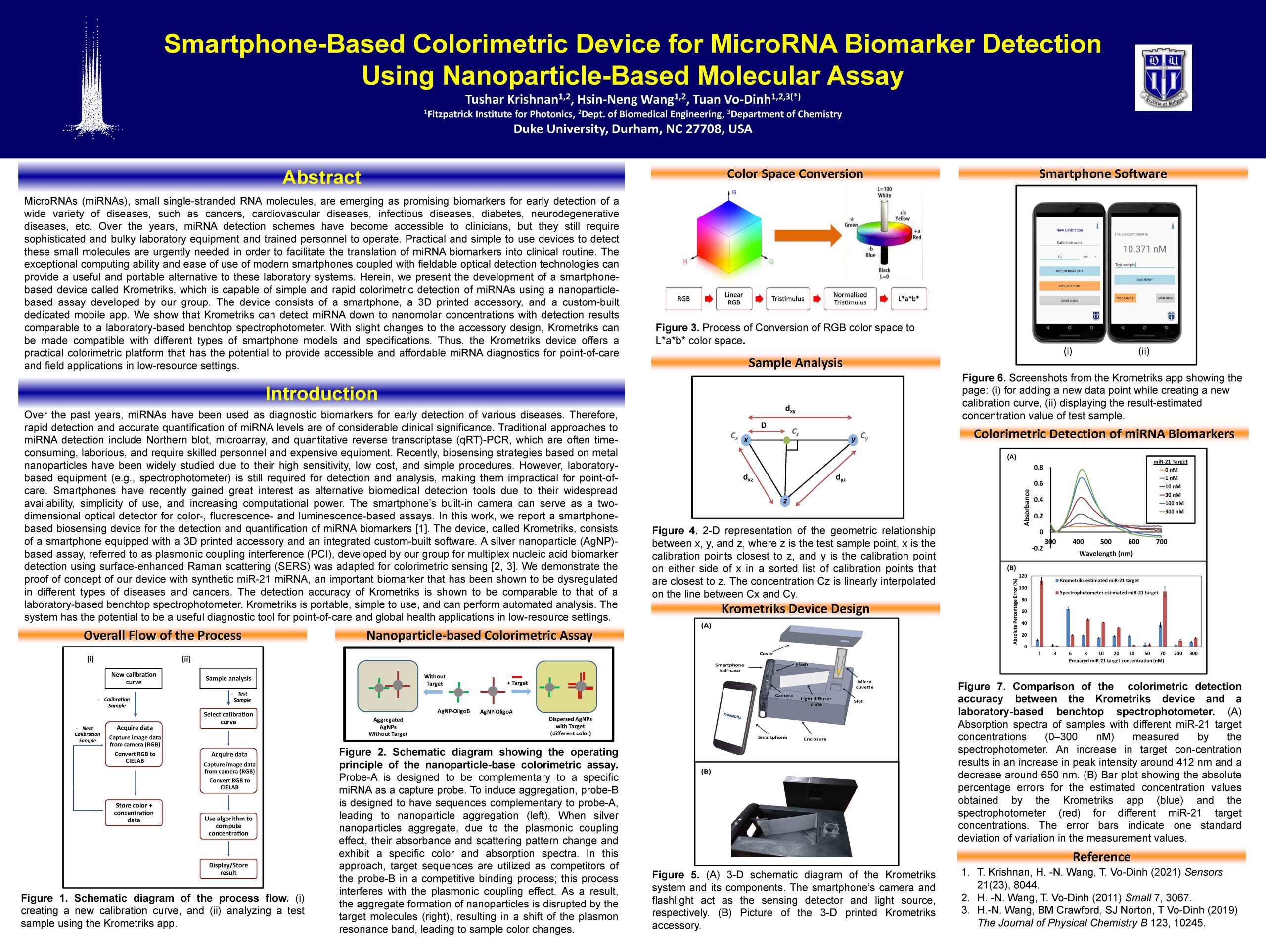
14. Smartphone-Based Colorimetric Device for MicroRNA Biomarker Detection Using Nanoparticle-Based Molecular Assay
Tushar Krishnan, Hsin-Neng Wang, Tuan Vo-Dinh
Department of Biomedical Engineering, Duke University
MicroRNAs (miRNAs), small single-stranded RNA molecules, are emerging as promising biomarkers for early detection of a wide variety of diseases, such as cancers, cardiovascular diseases, infectious diseases, diabetes, neurodegenerative diseases, etc. Over the years, miRNA detection schemes have become accessible to clinicians, but they still require sophisticated and bulky laboratory equipment and trained personnel to operate. Practical and simple to use devices to detect these small molecules are urgently needed in order to facilitate the translation of miRNA biomarkers into clinical routine. The exceptional computing ability and ease of use of modern smartphones coupled with fieldable optical detection technologies can provide a useful and portable alternative to these laboratory systems. Herein, we present the development of a smartphone-based device called Krometriks, which is capable of simple and rapid colorimetric detection of miRNAs using a nanoparticle-based assay developed by our group. The device consists of a smartphone, a 3D printed accessory, and a custom-built dedicated mobile app. We show that Krometriks can detect miRNA down to nanomolar concentrations with detection results comparable to a laboratory-based benchtop spectrophotometer. With slight changes to the accessory design, Krometriks can be made compatible with different types of smartphone models and specifications. Thus, the Krometriks device offers a practical colorimetric platform that has the potential to provide accessible and affordable miRNA diagnostics for point-of-care and field applications in low-resource settings.
hsinneng.wang@duke.edu
15. Automated Detection of Sarcoma Tissue in a Murine Model Using a Portable Endogenous Fluorescence Spectroscopy Device
Virtual Poster Presentation: Ashutosh Raman

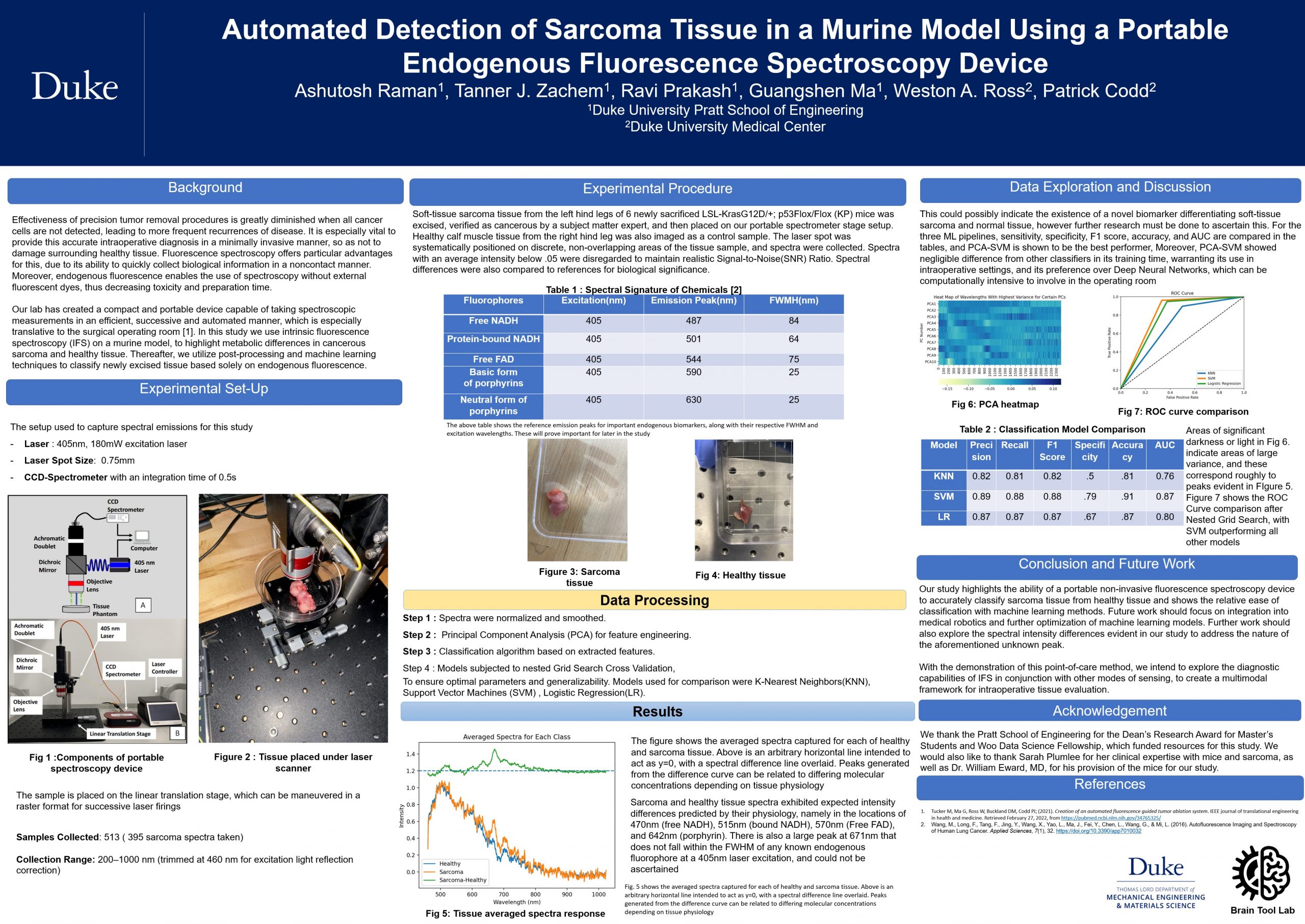
Ashutosh Raman, Tanner J. Zachem, Ravi Prakash, Guangshen Ma, Dr. Weston A. Ross, Dr. Patrick Codd
Department of Biomedical Engineering, Duke University; Department of Mechanical Engineering and Materials Science, Duke University; Duke University Medical Center, Duke University
Intro: Effectiveness of precision tumor removal procedures is greatly diminished when all cancer cells are not detected, leading to more frequent recurrences of disease. It is especially important to provide this accurate intraoperative diagnosis in a minimally invasive manner, so as not to damage surrounding healthy tissue. Fluorescence spectroscopy offers particular advantages for this, due to its ability to quickly collect biological information in a noncontact manner. Moreover, endogenous fluorescence enables the use of spectroscopy without external dyes, thus decreasing toxicity and preparation time. In this study we use intrinsic fluorescence spectroscopy (IFS) on a murine model, to highlight metabolic differences in cancerous sarcoma and healthy tissue. Thereafter, we utilize post-processing and machine learning techniques to classify newly excised tissue based solely on endogenous fluorescence. Methods: A 405nm, 180mW excitation laser with .75mm spot size and a CCD-Spectrometer with an integration time of .5s were used to capture spectral emissions for this study. Soft-tissue sarcoma tissue from the left hind legs of 6 newly sacrificed LSL-KrasG12D/+; p53Flox/Flox (KP) mice was excised, verified as cancerous by a subject matter expert, and then placed on our portable spectrometer stage setup. Healthy tissue from the right hind leg was also imaged as a control sample. The laser spot was systematically positioned on discrete, non-overlapping areas of the tissue sample, and spectra were collected. 513 samples were collected, with 395 sarcoma spectra taken. Wavelength collection ranges spanned from 200-1000nm, however, ranges were trimmed to 460-1000nm to correct for excitation light reflection. Spectra with an average intensity below .05 were disregarded to maintain realistic Signal-to-Noise Ratio. Lastly, spectra were normalized and smoothed. Principal Component Analysis (PCA) was used for feature engineering, followed by classification with K-Nearest Neighbors, Logistic Regression, Support Vector Machines, and a simple Artificial Neural Network (ANN). The former three were subjected to nested Grid Search Cross Validation, to ensure optimal parameters and generalizability. Results and Discussion: Sarcoma and healthy tissue spectra exhibited expected intensity differences predicted by their physiology, namely in the locations of 470nm (free NADH), and 570nm (Free FAD). Use of ANNs is substantiated, with accuracies around 94%, though their computational intensivity and training time remain prohibitive. For the three other ML pipelines, sensitivity, specificity, F1 score, accuracy, and AUC are compared, and PCA-SVM is shown to be the best performer, with testing accuracy of 88%, recall of 90%, and specificity of 83%. Moreover, PCA-SVM showed negligible difference from other classifiers in its training time, warranting its use in intraoperative settings, and its preference over ANNs. Conclusion/Future: Our study highlights the ability of a portable non-invasive fluorescence spectroscopy device to accurately classify sarcoma tissue from healthy tissue, and also shows the relative ease of classification with machine learning methods. Future work should focus on integration into medical robotics and further optimization of machine learning models. With the demonstration of this point-of-care method, we intend to explore the diagnostic capabilities of IFS in conjunction with other modes of sensing, to create a multimodal framework for intraoperative tissue evaluation.
ashutosh.raman@duke.edu
16. Spatial-Temporal Disorder Strength Measured by Quantitative Phase Imaging to Report Cell Stiffness
Virtual Poster Presentation: Steven Parker


Hillel B. Price, Steven Parker, Meghan Reynolds, Brenton D. Hoffman, Adam Wax
Quantitative phase imaging (QPI) has been shown to be a useful label-free method of quantifying nanometer scale cell information such as structure, dynamics, function, and physical properties. Optical volume (OV) has been shown to be a non-invasive way to measure cell physical properties like density. Disorder strength (LD ) measured via QPI can inform mechanical properties of cells like stiffness, related to Young’s modulus measured via atomic force microscopy. It is hypothesized that spatial-temporal measurements by QPI can identify when cells have received a stimulus, such as hypo-osmotic shock.
steven.m.parker@duke.edu
17. WeaVE, a Wearable Variable Emittance Device for Personal Thermoregulation
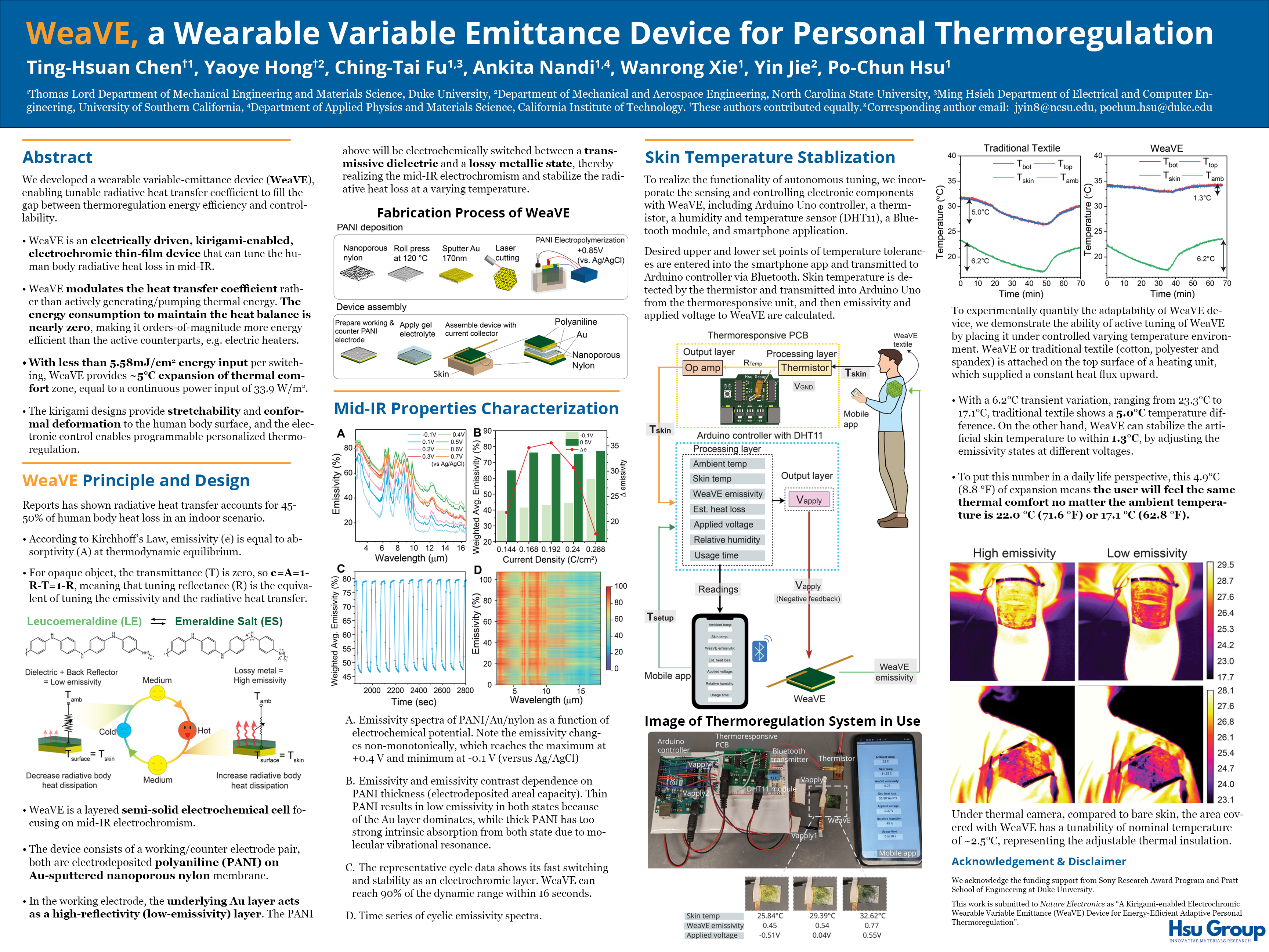
Ting-Hsuan Chen, Yaoye Hong, Ching-Tai Fu, Ankita Nandi, Wanrong Xie, Dr. Yin Jie, Dr. Po-Chun Hsu
Thomas Lord Department of Mechanical Engineering and Materials Science, Duke University
Department of Mechanical and Aerospace Engineering, North Carolina State University
Ming Hsieh Department of Electrical and Computer Engineering, University of Southern California
Department of Applied Physics and Materials Science, California Institute of Technology
For centuries, people have put effort to improve the thermal performance of clothing to adapt to varying temperatures. However, most clothing we wear today only offers a single-mode insulation. In this work, we developed a wearable variable-emittance device (WeaVE), enabling tunable radiative heat transfer coefficient to fill the missing gap between thermoregulation energy efficiency and controllability. WeaVE is an electrically driven, kirigami-enabled electrochromic thin-film device that can effectively tune the mid-infrared thermal radiation heat loss of the human body. The kirigami designs provide stretchability and conformal deformation attached to the human body surface, and the electronic control enables programmable personalized thermoregulation. With less than 5.58mJ/cm2 energy input per switching, WeaVE provides 5°C expansion of thermal comfort zone, which is equivalent to a continuous power input of 33.9 W/m2. This non-volatile characteristic substantially decreases the required energy while maintaining the on-demand controllability, thereby providing vast opportunities for the next generation of smart personal thermal managing fabrics and wearable technologies.
tc282@duke.edu
18. Growth Kinetics of Single Polymer Particles in Solution via Active-Feedback 3D Tracking

Donggeng Yu, Antonio Garcia lV, Suzanne A. Blum, Kevin D. Welsher
1. Department of Chemistry, Duke University, Durham, NC, USA
2. Department of Chemistry, University of California, Irvine, Irvine, CA, USA
The ability to directly observe chemical reactions at the single-molecule and single-particle level has enabled the discovery of behaviors otherwise obscured by the ensemble averaging in bulk measurements. However powerful, a common restriction of these studies to date has been the absolute requirement to surface tether or otherwise immobilize the chemical reagent/reaction of interest. This constraint arose from a fundamental limitation of conventional microscopy techniques, which could not track molecules or particles rapidly diffusing in three dimensions, as occurs in solution. However, much chemistry occurs in the solution phase, leaving single-particle/-molecule analysis of this critical area of science beyond the scope of available technology. Here we report the first solution-phase studies and measurements of any chemical reaction at single-particle/-molecule level in freely diffusing solution. During chemical reaction, freely diffusing polymer particles (D ~ 10-12 m2/s) yielded single-particle 3D trajectories and real-time volumetric images that were analyzed to extract the growth rates of individual particles. These volumetric images show that the average growth rate is a poor representation of the true underlying variability in polymer-particle growth behavior. These data revealed statistically significant populations of faster- and slower-growing particles at different depths in the sample, showing emergent heterogeneity while particles are still in the solution phase. These results go against the prevailing premise that chemical processes freely diffusing in solution will exhibit uniform kinetics. These new understandings of mechanisms behind polymer growth variations bring about an exciting opportunity to control particle-size and plausibly molecular weight polydispersity by the rational design of conditions to dictate spatial growth gradients. We anticipate that these studies will launch a new field of solution-phase, nonensemble-averaged measurements of chemical reactions.
dy66@duke.edu
19. Estimation of Spatial and Geometric Effects of Thick Tissue Imaging Using a GPU-Accelerated Monte Carlo Simulation Code
In Person Poster Presentation: Kyle Ferguson


Kyle S. Ferguson, Dr. Joel A. Greenberg
Medical Physics Graduate Program, Department of Electrical and Computer Engineering, Duke University
X-ray diffraction (XRD) imaging of thin biospecimen samples can provide rich information about various tissue’s presence and disease state. We have shown, for example, that XRD imaging can identify cancer in breast tissue with accuracy that exceeds current AI-based whole-slide imaging. We look to translate our successful prior studies in thin samples to thicker medical tissue samples for intra-operative intervention and diagnostic imaging. In thin samples, single-scatter events dominate, and solving the inverse problem of scatter localization is straightforward; however, as the sample thickness increases, multiple scatter and geometric blurring effects become important. To investigate the role of multiple scatter in unprepared biospecimen imaging, small animals, and potentially in-vivo applications, we utilize a Monte Carlo GPU-accelerated (MCGPU) photon transport simulation code. MC-GPU will enable us to explore how the energy and angular distribution of the multiple scatter signal depends on the properties of the incident beam and the composition of the specimen. In addition, we may extract information from multiple scatter that is useful, providing us with additional tissue specificity. In this poster, I will report on my work to date on performing a fundamental investigation into the role of multiple scatter and provide a framework for analyzing and implementing XRD imaging on thick samples.
kyle.ferguson@duke.edu
20. Multimodality X-ray transmission and 3D diffraction scanner for molecular analysis of cancer specimens
Virtual Poster Presentation: Zachary Gude


Zachary Gude, Joel Greenberg, Shannon J. McCall, Anuj J. Kapadia
Medical Physics Graduate Program, Duke University
Conventional X-ray transmission imaging techniques provide high spatial resolution but lack the molecular specificity often needed to differentiate between similarly attenuating tissues. Conventional X-ray diffraction (XRD) techniques can differentiate tissues at a molecular level, but typically requires destructive sample preparation and analyzes a sample at a single spatial location. Our group has developed a coded aperture based XRD system for thin (<1cm thick) samples that enables transverse (2D) XRD imaging of the scatter signal integrated throughout the sample depth. Additionally, it combines the XRD imaging capabilities with an X-ray transmission image to provide the user both high spatial resolution and molecular specificity We have previously shown that this multimodality system can identify cancer within mixed pathology samples with high accuracy. However, extending these techniques to thick samples by realizing depth resolution could enable new studies investigating the genesis and evolution of cancer. Our most recent work has been modifying the combined XRD & Transmission system and its data processing for the purpose of realizing 3D XRD imaging. In a system initially built for only 2D XRD, we have shown that 3D XRD is possible with a depth resolution of about 7 mm. This work helps validate our models and expectations as we move towards building a system optimized for volumetric XRD.
zwg@duke.edu
21. Accuracy of oxygen saturation measures from consumer wearable devices

Yihang Jiang, Biomedical Engineering Department, Duke University, Durham, NC, USA
Connor Spies, Palmer Lab, School of Medicine, Duke University, Durham, NC, USA
Will Wang, Biomedical Engineering Department, Duke University, Durham, NC, USA
Jessilyn P. Dunn PHD, Duke Clinical Research Institute, Departments of Biomedical Engineering and Bioinformatics & Biostatistics, Duke University, Durham, NC, USA
Satasuk Joy Bhosai MD MPH, Duke Clinical Research Institute, joint with Global Health Innovation Center, Department of Global Health
Laurie Snyder MD, Duke Clinical Research Institute, Department of Medicine
As COVID-19 has spread globally, the power of leveraging existing digital tools has become increasingly important. Remote tools built upon ubiquitous wearable device technologies can empower patients to monitor their oxygen (SpO2) at home conveniently. However, there has been no systematic evaluation to date on the accuracy and reliability of SpO2 monitoring from commercial wearable devices. We evaluated four consumer smartwatches that offer pulse oximetry, including the Apple Watch Series 7, Garmin Venu 2s, Garmin Fenix 6 Pro, and Withings ScanWatch and compared results with the reference device Masimo MightySat Rx.
yihang.jiang@duke.edu
© 2026 Frontiers in Photonics Science & Technology 2022
Theme by Anders Noren — Up ↑