A Virtual Symposium hosted by the Fitzpatrick Institute for Photonics | March 7-8, 2022

Special Themes
Photonics and Astronomy: A Quest Beyond the Stars

Photonics for Health in the Pandemic Era

Next-Generation Photonic Sensing and Imaging

logohope

Virtual Agenda

Introduction

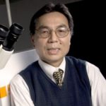
Tuan Vo-DinhTuan Vo-Dinh

Director of the Fitzpatrick Institute for Photonics,
R. Eugene and Susie E. Goodson Professor of Biomedical Engineering and Professor of Chemistry
Duke University

Dr. Vo-Dinh is R. Eugene and Susie E. Goodson Distinguished Professor of Biomedical Engineering, Professor of Chemistry, and Director of the Fitzpatrick Institute for Photonics at Duke University. His research interests involve the development of advanced technologies for the protection of the environment and the improvement of human health. His research activities are focused on nanophotonics, biophotonics, nano-biosensors, biochips, molecular spectroscopy, medical diagnostics and therapy, immunotherapeutics, bioenergy research, personalized medicine, and global health. Dr. Vo-Dinh has received seven R&D 100 Awards for Most Technologically Significant Advance in Research and Development for his pioneering research and inventions of innovative technologies. He has received the Gold Medal Award, Society for Applied Spectroscopy (1988); the Languedoc-Roussillon Award (France) (1989); the Scientist of the Year Award, ORNL (1992); the Thomas Jefferson Award, Martin Marietta Corporation (1992); two Awards for Excellence in Technology Transfer, Federal Laboratory Consortium (1995, 1986); the Inventor of the Year Award, Tennessee Inventors Association (1996); and the Lockheed Martin Technology Commercialization Award (1998), The Distinguished Inventors Award, UT-Battelle (2003), and the Distinguished Scientist of the Year Award, ORNL (2003). In 1997, Dr. Vo-Dinh was presented the prestigious Exceptional Services Award for distinguished contribution to a Healthy Citizenry from the U.S. Department of Energy. In 2011 Dr. Vo-Dinh received the Award for Spectrochemical Analysis from the American Chemical Society (ACS) Division of Analytical Chemistry. In 2019, Dr, Vo-Dinh was awarded the Sir George Stokes Award from the Royal Society of Chemistry (United Kingdom).

Welcome Remarks

Sally A. KornbluthSally A. Kornbluth

Duke University Provost
Jo Rae Wright Distinguished Professor
Duke University

Sally Kornbluth, Ph.D. was appointed Duke University Provost on July 1, 2014. Kornbluth served as Vice Dean for Basic Science at Duke University School of Medicine from 2006-2014. In this role, she served as a liaison between the Dean’s office and the Basic Science Department Chairs and faculty, including oversight of the biomedical graduate programs in the School of Medicine, implementation of programs to support the research mission of the basic science faculty, and oversight of new and existing core laboratories.

Kornbluth received a B.A. in Political Science from Williams College in 1982 and a B.S. in Genetics from Cambridge University, England in 1984 where she was a Herchel Smith Scholar at Emmanuel College. She received her Ph.D. from The Rockefeller University in 1989 in Molecular Oncology and went on to postdoctoral training at the University of California, San Diego. She joined the Duke faculty in 1994 and is currently the Jo Rae Wright University Professor.

Kornbluth’s research interests include the study of cell proliferation and programmed cell death, areas of central importance for understanding both carcinogenesis and degenerative disorders. She has published extensively in these areas, studying these problems in a variety of model organisms.

Dr. Jerome LynchJerome Lynch

Vinik Dean, Pratt School of Engineering
Professor of Civil and Environmental Engineering
Professor of Electrical and Computer Engineering
Duke University

Dr. Jerome Lynch is the Vinik Dean of Engineering of the Pratt School of Engineering at Duke University; he also holds the titles of Professor of Civil and Environmental Engineering and Professor of Electrical and Computer Engineering. Prior to joining Duke, he was a tenured faculty member of the University of Michigan from 2003 to 2021. Dr. Lynch completed his graduate studies at Stanford University where he received his Ph.D. in Civil and Environmental Engineering, M.S. in Civil and Environmental Engineering, and M.S. in Electrical Engineering. Dr. Lynch also received his B.E. in Civil and Environmental Engineering from the Cooper Union in New York City. He is a preeminent scholar working in the field of advanced sensing and information technologies for the monitoring and control of civil infrastructure systems. Among his many research achievements, he was the Director of the $19.2 million National Institute of Standards and Technology (NIST) Technology Innovation Program Center on Cyber-Enabled Wireless Monitoring Systems for the Protection of Deteriorating Infrastructure Systems from 2009 to 2014. In 2016, Dr. Lynch was appointed the Director of the University of Michigan Urban Collaboratory, a cross-campus research institute that facilitates close collaboration with city stakeholders to prototype solutions to community challenges using smart city technologies and socially engaged design methods. Dr. Lynch’s research and teaching accomplishments have been celebrated with several honors including the 2005 ONR Young Investigator Award, 2009 NSF CAREER Award, 2009 Presidential Early Career Award for Scientists and Engineers, 2012 American Society of Civil Engineering (ASCE) Leonardo da Vinci Award, and 2014 ASCE Huber Award.  He was elected Fellow of the Engineering Mechanics Institute in 2021.  Dr. Lynch is a seasoned entrepreneur having launched multiple high-tech companies including Civionics Inc. which was acquired in 2018, resulting in the establishment of Percev LLC as a subsidiary of Grace Technologies. 

Symposium Keynote

Andrea Ghez

2020 Nobel Laureate in Physics
Professor of Physics & Astronomy
University of California, Los Angeles
California, USA

Andrea M. Ghez, professor of Physics & Astronomy and Lauren B. Leichtman & Arthur E. Levine chair in Astrophysics, is one of the world’s leading experts in observational astrophysics and heads UCLA’s Galactic Center Group. Best known for her ground-breaking work on the center of our Galaxy, which has led to the best evidence to date for the existence of supermassive black holes, she has received numerous honors and awards including  the Nobel Prize in 2020, she became the fourth woman to be awarded the Nobel Prize in Physics, sharing one half of the prize with Reinhard Genzel (the other half of the prize being awarded to Roger Penrose). The Nobel Prize was awarded to Ghez and Genzel for their Independent discovery of a supermassive compact object, now generally recognized to be a black hole, in the Milky Way’s galactic center, the Crafoord Prize in Astronomy from the Royal Swedish Academy of Science (she is the first woman to receive a Crafoord prize in any field), Bakerian Medal from the Royal Society of London, a MacArthur Fellowship, election to the National Academy of Sciences, the American Academy of Arts & Sciences, and the American Philosophical Society.  


Her work on the orbits of stars at the center of the Milky Way has opened a new approach to studying black holes and her group is currently focused on using this approach to understand the physics of gravity near a black hole and the role that black holes plays in the formation and evolution of galaxies.

Advances in high resolution imaging technology enabled Ghez’s work and her group continues to work on pushing the frontiers of these technologies forward.   She serves on several leadership committees for the W. M. Keck Observatory, which hosts the largest telescopes in the world, and the future Thirty Meter Telescope.

Ghez is also very committed to the communication of science to the general public and inspiring young girls into science. Her work can be found in many public outlets including TED, NOVA’s Monster of the Milky Way, Discovery’s Swallowed by a Black Hole, TED, and Griffith Observatory.

Ghez earned her B.S from MIT in 1987, and her PhD from Caltech in 1992 and has been on the faculty at UCLA since 1994.

For more information see http://www.galacticcenter.astro.ucla.edu and http://www.astro.ucla.edu/~ghez


Learn about new developments in the study of supermassive black holes. Through the capture and analysis of twenty years of high-resolution imaging, the UCLA Galactic Center Group has moved the case for a supermassive black hole at the center of our galaxy from a possibility to a certainty and provided the best evidence to date for the existence of these truly exotic objects.  This was made possible with the first measurements of stellar orbits around a galactic nucleus. Further advances in state-of-the-art of high-resolution imaging technology on the world’s largest telescopes have greatly expanded the power of using stellar orbits to study black holes. Recent observations have revealed an environment around the black hole that is quite unexpected (young stars where there should be none; a lack of old stars where there should be many; and a puzzling new class of objects). Continued measurements of the motions of stars have solved many of the puzzles posed by these perplexing populations of stars. This work is providing insight into how black holes grow and the role that they play in regulating the growth of their host galaxies.  Measurements this past year of stellar orbits at the Galactic Center have provided new insight on how gravity works near a supermassive hole, a new and unexplored regime for this fundamental force of nature.

“From the Possibility to the Certainty of a Supermassive Black Hole”

FIP Pioneer Award Presentation

2022 Winner: Andrea Gehz

2020 Nobel Laureate in Physics
Professor of Physics & Astronomy
University of California, Los Angeles
California, USA

To recognize the dedicated contributions that researchers have made in the photonics community, the Fitzpatrick Institute for Photonics (FIP) created the FIP Pioneer Award to honor these individuals.

Past Winners
  • 2021 – Rainer Weiss
  • 2020 – Donna Strickland
  • 2019 – Shuji Nakamura
  • 2018 – Steven Chu
  • 2017 – Eric Betzig
  • 2016 – W.E. Moerner
  • 2015 – Theodor W. Hänsch
  • 2014 – Roger Y. Tsien
  • 2013 – William D. Phillips
  • 2011 – Martin Chalfie
  • 2010 – Ahmed H. Zewail
  • 2008 – John L. Hall
  • 2006 – Charles Townes

Pioneer Award Presentation by FIP Director Tuan Vo-Dinh
and Message from
Awardee Dr. Andrea Gehz

Plenary LectureS

Duncan Graham

Distinguished Professor, Head of Department
Department of Pure and Applied Chemistry
Technology and Innovation Centre
University of Strathclyde
Glasgow, Scotland

Duncan Graham is a Distinguished Professor, Associate Principal and Executive Dean of the Faculty of Science at the University of Strathclyde in Glasgow.  He obtained his BSc Honours (1992) and PhD in Chemistry (1995) from the University of Edinburgh before joining the University of Strathclyde in 1996.  In 2007 he was elected to the Royal Society of Edinburgh then awarded the RSC’s Corday Morgan prize in 2009, a Royal Society Wolfson Merit Award in 2010 and the RSC’s Theophilus Redwood award in 2016.  He served as Editor in Chief of the RSC journal Analyst for 7 years and serves on several editorial advisory boards including Chemical Society Reviews and Chemical Science.  He was president of the analytical division of the Royal Society of Chemistry (2017-2020), and chair of the analytical chemistry trust fund (2017-2020).  He is currently a trustee of and chair of the Publishing Board for the RSC (2020-2024).  His scientific interests are in using synthetic chemistry to produce sensors that respond to a specific biological species or events as measured by Raman spectroscopy or SERS and collaborating with scientists from different disciplines to exploit these approaches. 

Raman spectroscopy is an attractive technique for the analysis of biomolecules due to the rich information provided.  However, due to the lack of sensitivity, Raman spectroscopy has struggled to become adopted in many widespread biological applications.  To enhance the sensitivity of Raman spectroscopy, surface enhanced Raman spectroscopy (SERS) can be used and this presentation will focus on the latest developments of using SERS from Strathclyde.  In the first example, a new lateral flow based assay for the sensitive detection of biomarkers for infectious disease will be presented.  Following this a new observation on the nature of the metallic substrate used to provide enhancement of Raman scattering will be presented where an unusual temperature dependence on the enhancement of the Raman scattering has been found.  This is an interesting phenomenon where data will be presented on a new optical phenomenon which has been discovered in our laboratories resulting in an increase in SERS intensity due to plasmonic heating of hollow gold nanoshells.  The second part of the presentation will focus on the use of stimulated Raman scattering to provide rapid, highly informative images of multiple live single cells and how they respond to different drug treatments.  Phasor analysis of the data allows greater discrimination between the different regions within the cell moving towards sensitive quantitative analysis of a phenotypical state of these live cells.  Examples will include the use of alkyne tagged Raman imaging approaches where specific molecules have been synthesized to report on cellular conditions such as pH and also how they have been designed to locate in specific sub cellular compartments.  Overall the two areas for discussion in this presentation will highlight the latest developments in our laboratory in terms of using advanced Raman scattering techniques for the analysis of biological systems and biomolecules.


“Raman, SERS and SRS Analysis of Biomolecules”

Dr. Thomas ThundatThomas Thundat

Professor
Empire Innovation Professor
Chemical and Biological Engineering
School of Engineering and Applied Sciences
University of Buffalo, New York, USA

Dr. Thomas Thundat is an Empire Innovation professor in the Department of Chemical and Biological Engineering at the University at Buffalo, The State University of New York, Buffalo, NY. He is also Distinguished Professor (honorary) at the Indian Institute of Technology, Madras.  He was a Canada Excellence Research Chair professor at the University of Alberta, Edmonton, Canada (2010-2017). He was a research group leader for Nanoscale Science and Devices Group, and a UT-Battelle Corporate Fellow of the Oak Ridge National Laboratory (ORNL) until 2010. He received his Ph.D. in physics from the State University of New York at Albany. He is the author of over 450 publications in refereed journals, 85 conference proceedings, 45 book chapters, and 45 US patents. He is an elected Fellow of the American Physical Society (APS), the Electrochemical Society (ECS), the American Association for Advancement of Science (AAAS), the American Society of Mechanical Engineers (ASME), the SPIE, American Society of Biomedical and Biological Engineers (AIMBE), Institute of Electrical and Electronics Engineers (IEEE), and the National Academy of Inventors (NAI). His research is currently focused on developing new concepts in nanomechanical sensing, energy conversion, electrical energy transmission using single wire, and novel concepts for charge separation and storage.

Resonant optical excitation of targeted molecules provides very high spectroscopic selectivity in chemical and biomolecular detection.  Although radiative relaxation is often used for chemical and biological sensing, obtaining high sensitivity and selectivity in detection requires complex and bulky equipment.  However, non-radiative relaxation of resonantly excited molecules, that can generate extremely small amounts of heat by photothermal effect, can be detected using microfabricated cantilevers. Cantilevers, when fabricated as bi-material beams, have a thermal sensitivity in the milli Kelvin range at ambient temperature. In addition to photothermal signatures of adsorbed molecules, micromechanical resonators can also provide adsorbed mass and adsorption energy with a very high sensitivity. These cantilever resonators can also be fabricated as microfluidic channels for characterizing confined or flowing liquid samples in them. This approach provides a label-free and receptor-free method for molecular recognition, and overcomes many of the selectivity challenges encountered when using receptor-based approaches. Understanding energy dissipation at resonance can impart additional information for enhancing the selectivity. Multi-modal, multi-physics data obtained with the nanomechanical platform, when analyzed using deep learning techniques, can enhance the selectivity, sensitivity, and reliability even in complex mixtures and environments. I will discuss examples of the detection of drugs, interaction of drugs with drug-resistant bacteria and the unravelling of DNA, etc. in this presentation.

“Molecular Recognition Using Nanomechanical Photothermal Effects”

Invited Lectures

Hatice Altug

Professor, Department of Bioengineering
Head of Bionanophotonic Systems Laboratory
Ecole Polytechnique Fédérale de Lausanne (EPFL)
Lausanne, Switzerland

Hatice Altug is full professor in the Institute of Bioengineering at Ecole Polytechnique Federale de Lausanne (EPFL), Switzerland. She is also the director of EPFL Doctoral School in Photonics. Between 2007 and 2013, she was professor in the Electrical and Computer Engineering Department at Boston University, U.S. She received her Ph.D. in Applied Physics from Stanford University (U.S.) in 2007. She received her B.S. in Physics from Bilkent University (Turkey) in 2000.

Prof. Altug is the recipient of the U.S. Presidential Early Career Award for Scientists and Engineers in 2011, which is the highest honor bestowed by the United States government on outstanding scientists and engineers in their early career,  Optical Society of America Adolph Lomb Medal, which is presented to a person who has made a noteworthy contribution to optics at an early career stage, and the European Physical Society (EPS) Emmy Noether Distinction. She received the European Research Council (ERC) Consolidator Grant, ERC Proof of Concept Grant, U.S. Office of Naval Research Young Investigator Award, U.S. National Science Foundation CAREER Award, Massachusetts Life Science Center New Investigator Award, IEEE Photonics Society Young Investigator Award. She is an elected fellow of the Optical Society of America. She won Inventors’ Challenge competition of Silicon Valley in 2005. She has been named to Popular Science Magazine’s “Brilliant 10” list in 2011.

Prof. Altug is a leading expert in the fields of nanophotonics and its application to biosensing, spectroscopy and bioimaging. Her research has a broad coverage from optics, microfluidics, micro/nanofabrication, biochemistry to data science. Her laboratory introduces next-generation bianalytical technologies for label-free, real-time, and high-throughput analysis on biomolecules, pathogens and living systems for applications in life science research, disease diagnostics and point-of-care testing.


Emerging healthcare needs with global health crisis, personalized medicine and point-of-care diagnostics are demanding breakthrough developments in biosensing and bioanalytical tools. Current biosensors are lacking precision, bulky, and costly, as well as they require long detection times, sophisticated infrastructure and trained personnel, which limit their applications. My laboratory is focused on to address these challenges by exploiting novel optical phenomena at nanoscale and engineering toolkits such as nanophotonics, nanofabrication, microfluidics and data science. In particular, we use photonic nanostructures based on plasmonic and dielectric metasurfaces that can confine light below the fundamental diffraction limit and generate strong electromagnetic fields in nanometric volumes for new device functionalities. We develop new nanofabrication procedures that can enable high-throughput and low-cost manufacturing of nanophotonic biochips. We integrate our sensors with microfluidics for efficient analyte control. We combine smart data science methods with bioimaging and biosensing architectures to achieve unprecedented performance. In this talk I will present some of our recent work [1-15]. For example, I will introduce ultra-sensitive Mid-IR biosensors based on surface enhanced infrared spectroscopy for chemical specific detection of molecules, large-area chemical imaging and real-time monitoring of protein conformations in aqueous environment. I will describe portable, rapid and low-cost microarrays and their use for early disease diagnostics in real-world settings. I will also describe optofluidic biosensors that can perform one-of-a-kind measurements on live cells down to the single cell level, and provide their overall prospects in biomedical and clinical applications.

References:

[1] Leitis et al. “Wafer-Scale Functional Metasurfaces for Mid-Infrared Photonics and Biosensing”, Advanced Materials, Vol. 33, 2102232 (2021). 

[2] Cachot et al. “Tumor-Specific Cytolytic CD4 T Cells Mediate Immunity Against Human Cancer”, Science Advances, Vol. 7, eabe3348, (2021).

[3] John-Herpin et al. “Infrared Metasurface Augmented by Deep Learning for Monitoring Dynamics between All Major Classes of Biomolecules”, Advanced Materials, Vol. 33, 2006054 (2021)

[4] Jahani et al. “Imaging-Based Spectrometer-less Optofluidic Biosensor Based on Dielectric Metasurfaces for Detecting Extracellular Vesicles”, Nature Communications, Vol. 12, 3246 (2021).

[5] Tseng et al. “Dielectric Metasurfaces Enabling Advanced Optical Biosensors”, ACS Photonics, Vol. 8, p. 47-60, (2021). 

[6] Oh and Altug “Performance Metrics and Enabling Technologies for Nanoplasmonic Biosensors”, Nature Communications Vol. 9, p. 5263 (2018).

[7] Beluskin et al. “Rapid and digital detection of inflammatory biomarkers enabled by a novel portable nanoplasmonic imager” Small Vol. 16, 1906108 (2020).

[8] Yesilkoy et al. “Ultrasensitive Hyperspectral Sensing Based on High-Q Dielectric Metasurfaces”, Nature Photonics Vol. 13 p. 390-396 (2019).

[9] Gupta et al. “Self-assembly of nanostructured glass metasurfaces via templated fluid instabilities” Nature Nanotechnology Vol. 14, p. 320-327 (2019). 

[10] Mohammadi et al. “Accesible superchiral near-fields driven by tailored electric and magnetic resonances in all-dielectric nanostructures” ACS Photonics Vol. 6, p. 1939-1946 (2019).

[11] Tittl et al. “Imaging-Based Molecular Barcoding with Pixelated Dielectric Metasurfaces”, Science Vol. 360, p. 1105-1109 (2018).

[12] Etezadi et al. “Real-Time in-Situ Secondary Structure Analysis of Protein Monolayer with Mid-Infrared Plasmonic Nanoantennas”, ACS Sensors Vol. 3, p. 1109-1117 (2018).

[13] Li et al. “Label‐Free Optofluidic Nanobiosensor Enables Real‐Time Analysis of Single‐Cell Cytokine Secretion”, Small Vol. 14, 1870119 (2018).

[14] Soler et al. “Two-Dimensional Label-Free Affinity Analysis of Tumor Specific CD8 T Cells with a Biomimetic Plasmonic Sensor”, ACS Sensors Vol. 3, p. 2286-2295 (2018).

[15] Rodrigo et al. “Mid-Infrared Plasmonic Biosensing with Graphene” Science Vol 349, p. 165-168 (2015).


“Frontiers in Nanophotonics: Enabling Technology for Optical Biosensing and Bioimaging”

Dr. Joerg BewersdorfJoerg Bewersdorf

Professor, Departments of Cell Biology and
Biomedical Engineering
Yale University
New Haven, CT, USA

Joerg Bewersdorf is a Professor of Cell Biology and of Biomedical Engineering at Yale University. He received his Master’s degree (Dipl. Phys., 1998) and his doctoral degree in physics (Dr. rer. nat., 2002) training with Dr. Stefan W. Hell at the Max Planck Institute for Biophysical Chemistry in Goettingen, Germany. After 4 years at The Jackson Laboratory in Bar Harbor, Maine, he relocated his research group to Yale University in 2009. An optical physicist/biophysicist by training, Dr. Bewersdorf has been a long-time contributor to the field of super-resolution light microscopy development and the application of these techniques to cell biological questions.

Super-resolution optical microscopy has become a powerful tool to study the nanoscale spatial distribution of proteins of interest in cells over the last years. Imaging these distributions in the context of other proteins or the general cellular context is, however, still challenging. I will present recent developments of our lab which tackle this challenge: 4Pi-SMS microscopy simultaneously localizes up to three species of proteins with sub-10 nm localization precision in 3D [1]. A new fluorogenic DNA-PAINT probe enables fast, 3D whole-cell imaging without the need for optical sectioning, adding a versatile tool to the toolbox of single-molecule super-resolution probes [2]. Labeling proteins and other cellular components in bulk in our recent pan-Expansion Microscopy method provides ultrastructural context to the nanoscale organization of proteins, replacing complex correlative light/electron microscopy by an all-optical imaging approach [3].  

Financial Interest Disclosure: J.B. has financial interest in Bruker Corp. and Hamamatsu Photonics and is co-founder of a startup company related to Expansion Microscopy. 

[1] Zhang, Y., Schroeder, L.K., et al. “Fluorogenic probe for fast 3D whole-cell DNA-PAINT”. Nature Methods (2020). https://doi.org/10.1038/s41592-019-0676-4  

[2] Chung, K.K.H. et al. “Fluorogenic probe for fast 3D whole-cell DNA-PAINT”. bioRxiv (2020). https://doi.org/10.1101/2020.04.29.066886

[3] M’Saad, O., Bewersdorf, J. Light microscopy of proteins in their ultrastructural context. Nat Commun 11, 3850 (2020). https://doi.org/10.1038/s41467-020-17523-8

“All-optical Super-resolution Imaging of Molecules in Their Nanoscale Cellular Context”

Dr. Anna FrebelAnna Frebel

Associate Professor of Physics
Principal Investigator, MIT Kavli Institute for Astrophysics and Space Research
Massachusetts Institute of Technology
Boston, Massachusetts, USA

Anna Frebel is an Associate Professor in the Astrophysics Division of the the Physics Department at MIT. Originally from Germany, she received her PhD from the Australian National University’s Mt. Stromlo Observatory in 2007.  Following postdoctoral work at the University of Texas at Austin and the Harvard-Smithsonian Center for Astrophysics, Frebel joined the MIT physics faculty in 2012. She is a world leading expert in the fields of stellar archaeology and near-field cosmology which concerns the study of ancient 13 billion year old stars to understand the physical and chemical conditions of the early universe, the origin of the elements, and the formation of the Milky Way galaxy. For her research into the oldest stars in the universe and the early evolution of the chemical
elements she received awards such as the 2007 Charlene Heisler Prize
(Astronomical Society of Australia), the 2009 Ludwig-Biermann young astronomer award (German Astronomical Society) and the 2010 Annie Jump Cannon Award (American Astronomical Society). In 2013 she received a CAREER Award from the National Science Foundation, in 2016 she was named one of ScienceNews Magazine’s 10 scientists to watch. Frebel also enjoys communicating science to the public, e.g., through her popular science book “Searching for the oldest stars: Ancient Relics from the Early Universe” (also available in German “Auf der Suche nach den ältesten Sternen, S. Fischerverlag).

The first stars and first galaxies formed a few hundred million years after the Big Bang. Their emergence transformed the universe: the first heavy elements changed the gas physics and high energy photons reionized their surroundings. Hence, understanding this early era is at the frontier of modern astrophysics and cosmology. It can be well probed with 13 billion year old stars that are found in the outskirts of the Milky Way as well as its satellite dwarf galaxies.

Since their chemical composition has remained unchanged since their birth, an elemental abundance analyses of these stars reveals the nature of the first nucleosynthesis events within early star forming clouds soon after the Big Bang. The required high-resolution optical spectroscopic data is obtained with the 6.5 meter Magellan telescope in Chile.

“Discovering the oldest stars in the Milky Way and its dwarf galaxies
with high-resolution optical spectroscopy”

Dr. Sylvan GignanSylvain Gigan

Professor of Physics
Laboratoire Kastler-Brossel
Sorbonne University
Paris, France

Sylvain Gigan is Professor of Physics at Sorbonne Université in Paris, and group leader in Laboratoire Kastler-Brossel, at Ecole Normale Supérieure (ENS, Paris). His research interests range from fundamental investigations of light propagation in complex media, biomedical imaging, computational imaging, signal processing, to quantum optics and quantum information in complex media. He is also the cofounder of a spin-off: LightOn (www.lighton.ai) aiming at performing optical computing for machine learning and Big Data.

Light propagation in complex media, such as paint, clouds, or biological tissues, is a very challenging phenomenon, encompassing fundamental aspects in  mesoscopic and statistical physics. It is also of utmost applied interest, in particular for imaging. Wavefront shaping has revolutionized the ability to image through or in complex media. I will discuss how computational tools and machine learning allows to develop further wavefront shaping for imaging applications, and conversely discuss how the same complexity can be leveraged for optical computing tasks.

“A sneak peek with light into opaque materials : from imaging to computing”

Dr. Kimberly Hamad-SchifferliKimberly Hamad-Schifferli

Associate Professor, Department of Engineering
Affiliate Faculty, School for the Environment
University of Massachusetts Boston
Boston, Massachusetts, USA

Kimberly Hamad-Schifferli is an Associate Professor in the Department of Engineering and the School for the Environment at University of Massachusetts Boston. She obtained her S.B. in Chemistry from MIT in 1994 and Ph.D. in Chemistry from the University of California at Berkeley in 2000. She was a faculty member at MIT in the Department of Mechanical Engineering and the Department of Biological Engineering as a faculty member from 2002-2012. From 2012-2015 she was at MIT Lincoln Laboratory in the Bioengineering Systems and Technologies Group. Since 2015 she has been a faculty member at UMass Boston. She received an ONR Young Investigator Award and was named a Fellow of the Foresight Institute in 2017. Her research is focused on using nanotechnology for biological applications, such as infectious disease, cancer therapy, and food safety. Projects in her research lab include the development of low-cost diagnostics for viral diseases such as dengue, zika, yellow fever, and Ebola, the bacterial pathogen Vibrio parahaemolyticus, and biomarkers that can distinguish between viral and bacterial infections.

The global COVID-19 pandemic has underscored the need for innovations in disease diagnostics. The convergence of the fields of nanotechnology and medicine has resulted in new approaches for novel disease therapies, biomedical imaging and sensing, and numerous others. In particular, the use of gold nanoparticles in rapid diagnostics for infectious diseases has been emerging as an application with the potential to address some of the major challenges in global health. These assays are low-cost and can be used in rugged environments, so they are attractive for widespread deployment for disease surveillance, quarantining, and treatment.  Readout by eye is made possible by the gold nanoparticle-antibody conjugates, which have a strong absorption due to the nanoparticle surface plasmon resonance, thus providing sample-to-answer times of minutes. One opportunity for extending the capabilities of paper immunoassays that are not possible with traditional paper immunoassays lies in exploiting the unique size and material dependent properties of the nanoparticles. We describe a route for leveraging the optical properties of gold nanostars to use paper immunoassays for multiplexed diagnostics for yellow fever, dengue, and zika viruses. By adapting immunoassays for selective sensing as opposed to specific sensing and using machine learning of the color test lines, we are able to construct an assay for yellow fever non structural protein 1(NS1) using cross-reactive antibodies raised for dengue and zika. In addition, we discuss routes to increase the sensitivity of paper-based immunoassays via surface enhanced Raman spectroscopy (SERS). By using gold nanostars for Raman nanotags with different Raman reporter molecules, we can construct multiplexed assays for zika and dengue. We also discuss challenges associated with the biotic-abiotic interface in paper based immunoassays, which result in undesirable side effects such as non-specific adsorption and false positives. 

“Rapid Diagnostics for Infectious Diseases Using Gold Nanoparticles”

Dr. Catherine Heymans

Catherine Heymans

Professor, Astronomer Royal of Scotland
Institute of Astronomy
University of Edinburgh
Edinburgh, Scotland


Catherine Heymans is the Astronomer Royal for Scotland, Professor of Astrophysics at the University of Edinburgh and Director of the GCCL Institute at the Ruhr-Universität Bochum Germany.   She is well known for her research using deep sky observations to map the invisible dark matter and mysterious dark energy in our Universe, as recognised by a Max-Planck Humboldt Research Award and the Royal Astronomical Society Darwin Lectureship.  She is an enthusiastic science communicator, joining art, music, comedy, philosophy and science festivals, and is a regular contributor to BBC radio and television.

The Planck space mission has released exquisite observations of the early universe, providing the strongest evidence yet that the universe we live in is very dark indeed. Its precise results show that our universe is composed of 26.6% dark matter and 68.4% dark energy, while less than 5% is made up of the baryonic material that we are familiar with on Earth. With their long-standing quest to make these precision measurements essentially now concluded, cosmologists are rapidly turning their attention to a much bigger and further-reaching question: what is the exact nature of this dark universe? I will introduce the new directions being taken in Cosmology to map out the invisible dark matter and confront theories on the origins of dark energy. Interestingly the increasing precision recently reported in these late-time cosmological measurements reveals tension with Planck’s initial conclusions. Is this a sign that new data challenges lie ahead, or is it our first hint that the universe is truly exotic and that in order to understand the dark universe we will need some new physics that will forever change our cosmic view.

“New directions in Cosmology”

Dr. Laura LechugaLaura M. Lechuga

Professor, Group Leader
NanoBiosensors & Bioanalytical Applications Group
Catalan Institute of Nanoscience and Nanotechnology
Bellaterra, Barcelona, Spain

Prof. Laura M. Lechuga is Full Professor of the Spanish National Research Council (CSIC) and Head of the Nanobiosensors and Bioanalytical Applications Group at the Catalan Institute of Nanoscience and Nanotechnology (ICN2) in Barcelona (Spain) and at the Networking Biomedical Research Center (CIBER-BBN).   She has published over 260 articles, book chapters and proceedings, has 8 families of patents, and has presented her work worldwide in more than 365 invited talks. She has co-founded two spin-offs companies. The quality of her research has been recognised by prestigious prizes and awards, as among others, the Physics, Innovation and Technology Prize from the Spanish Royal Physics Society (RSEF) and BBVA Foundation (2016), the Ada Byron Prize (2020), the King Jaume I award in New Technologies and the Spanish National Research Prize in 2020, and the Burdinola Research award in 2021. From April 2020 to September 2021, she belonged to the Expert Scientific Panel advising the Ministry of Science and Innovation and the Spanish Government in the management of the COVID-19 pandemic.

COVID-19 pandemics has evidenced the urgent need for reliable and portable diagnostic tools, able to deliver rapid testing and screening of the population while providing sensitivity and specificity levels comparable to laboratory techniques. Biosensor technology is the most suitable one to tackle the challenging goal of offering fast and user-friendly diagnostics tests than can be employed at the point-of-need. In particular, photonic biosensors can provide sensitive, reliable and selective analysis, while reducing the time-to-result, decreasing and/or eliminating sample transport, and using low sample volume. And, more importantly, photonic biosensor technology can provide quantification, for example, of the SARS-CoV-2 viral load.

Our group have demonstrated cutting-edge nanophotonic biosensor point-of-care (PoC) devices (as the Bimodal Waveguide Interferometer) that enables ultrasensitive analysis of body fluids in few minutes and in decentralized settings. We have demonstrated sensitivities at the fM-aM level for clinical biomarkers diagnostics, as for example the direct specific miRNA detection in urine and the whole pathogen detection directly in human samples. During the pandemic, we have been working in the EU project CoNVat, one of the first diagnostics projects funded by the H2020 EU program to fight against COVID-19. Our biosensor technology can provide a rapid immunoassay detection (less than 20 minutes) of whole units of SARS-CoV-2 without the need of PCR or other time-consuming treatments with an outstanding sensitivity of around 100 FFU/mL in a single-step immunoassay. In addition, we have developed a quantitative serological biosensor with exceptional limits of detection, demonstrating a sensitivity of 99% and specificity of 100%, outperforming available techniques like immunoassays or rapid tests.

“Nanophotonics Biosensors for ultrasensitive diagnostics at the Point-of Need”

Dr. Dan OronDan Oron

Professor
The Harry Weinrebe Professional of Laser Physics
Weizmann Institute of Science
Rehovot, Israel

Dan Oron earned a B.Sc. in mathematics and physics from the Hebrew university in 1994. He earned his M.Sc. degree in physics (working on hydrodynamic instability) from Ben-Gurion University of the Negev in 1998 and received his Ph.D., also in physics, from the Weizmann Institute of Science in 2005, under the guidance of Prof. Yaron Silberberg. After conducting postgraduate research with Prof. Uri Banin at the Hebrew University for two years, he joined the staff of the Weizmann Institute in April 2007. He is currently a professor at the department of Molecular Chemistry and Materials Science at the Weizmann institute. His main research interests are at the interface between light and the nanoscale, studying both the interaction of light with nanostructured materials (mostly inorganic and hybrid semiconductor nanocrystals), optical superresolution methods harnessing both quantum and classical fluctuations in light emission and the optics of biological nanostructured materials.

Far-field optical microscopy beyond the Abbe diffraction limit, making use of nonlinear excitation (e.g. STED), or temporal fluctuations in fluorescence (PALM, STORM, SOFI) is already a reality. In contrast, overcoming the diffraction limit using non-classical properties of light is very difficult to achieve due to the difficulty in generating quantum states of light and their inherent fragility. Here, we experimentally demonstrate practical superresolution microscopy based on quantum properties of light naturally emitted by fluorophores used as markers in fluorescence microscopy. Our approach is based on photon antibunching, the tendency of fluorophores to emit photons one by one rather than in bursts.
Since the non-classical intensity correlations carry higher spatial frequency information, they can be utilized to enhance image resolution. We demonstrate how antibunching can improve the resolution capabilities of image-scanning confocal microscopy in all three dimensions1, and show how the synthesis of classical and quantum information enables us to apply algorithmic resolution augmentation methods2. Finally, we show that these methods are compatible with currently developed SPAD arrays, serving as small single-photon imaging detectors, and thus require little infrastructure investment3.

Notably, the use of photon antibunching readily lends itself to quantitative imaging modalities since the degree of antibunching is intimately related to the emitter density. Potential approaches to such quantitative superresolution microscopy using a simple confocal microscope setup will be discussed.

References
[1] R. Tenne et al., “Super-resolution enhancement by quantum image scanning microscopy”, Nature Photonics 13, 116 (2019).
[2] U. Rossman et al., “Rapid quantum image scanning microscopy by joint sparse reconstruction”, Optica 6, 1290 (2019).
[3] G. Lubin et al., “Quantum correlation measurement with single photon avalanche diode arrays”, Optics Express 27, 32863 (2019).

“Quantum enhanced superresolution microscopy”

Feryal Özel

Professor
Departments of Astronomy and Physics
University of Arizona, Tuscson, Arizona, USA

Feryal Ozel is a professor of Astronomy and Physics at the University of Arizona. She received her BS from Columbia University, her MS from the Niels Bohr Institute in Copenhagen, and her PhD from Harvard University. She was a NASA Hubble fellow at the Institute for Advanced Study at Princeton. Dr. Ozel pioneered the measurements of neutron star radii, which provided new constraints on the equation of state of cold ultra-dense matter. She also carried out the first simulations of the images of low-luminosity black holes that guided the development of interferometric imaging efforts from the X-rays to the Event Horizon Telescope at mm-wavelengths. She is a member of the Event Horizon Telescope Science Council and has been the lead of the modeling and analysis working group. She was also the co-chair of NASA’s Next Generation Mission Concept Study for Lynx X-ray Observatory and has served as the chair of NASA’s Astrophysics Advisory Committee. She is a Fellow of the American Physical Society. In recognition of her work, she has received numerous awards including the APS Maria Goeppert Mayer award, the Guggenheim Fellowship, the Radcliffe Fellowship and, with the EHT collaboration, the Breakthrough Prize, the Rossi Prize of the AAS, and the Diamond Achievement award from the National Science Foundation.

The Event Horizon Telescope recently imaged the supermassive black hole in the center of the M87 galaxy. I will describe the experiment and the methods used to obtain the image. I will also describe the extensive computational modeling effort we have undertaken to interpret the observations. I will finally show how we used this measurement to infer the the properties of the black hole and test the theory of of General Relativity. 

“From Images of a Black Hole to Tests of General Relativity”

Dr. Zac SchultzZachary Schultz

Associate Professor
Department of Chemistry and Biochemistry
The Ohio State University
Columbus, Ohio, USA

Zachary D. Schultz, Ph.D., is an associate professor at The Ohio State University. Prof. Schultz earned his B.S. degree from the Ohio State University in 2000 and Ph.D. from the University of Illinois at Urbana-Champaign in 2005. As a graduate student, he was recognized with an ACS Division of Analytical Chemistry Graduate Fellowship (2004). Upon completing his Ph.D., he was awarded a National Research Council Postdoctoral Fellowship to conduct research at the National Institute of Standards and Technology (USA). His research at NIST was performed largely in collaboration with Ira Levin at the National Institutes of Health (USA). Following his postdoctoral training at NIST, Dr. Schultz continued as a research fellow with Dr. Levin using vibrational spectroscopy and microscopy to study biomembrane systems. While at the NIH, Dr. Schultz was awarded an NIH Pathway to Independence Award. Dr. Schultz began his independent career as an assistant professor of chemistry and biochemistry at the University of Notre Dame in 2009 and was promoted with tenure to associate professor in 2015. In January of 2018, Prof. Schultz moved his research program to Ohio State.  Prof. Schultz has served on the Analytical Chemistry Editorial Advisory Board’s Features Panel and is currently on the Editorial Advisory board of Luminescence (Wiley), Analytical Methods (RSC), and is joining the EAB for The Journal of Physical Chemistry A/B/C in 2022. Prof. Schultz was recognized as a Cottrell Scholar in 2013, elected a Fellow of the American Association for the Advancement of Science (AAAS) in 2019, and was awarded the Craver Award for applied vibrational spectroscopy from the Coblentz Society in 2021. Prof. Schultz’s research focuses on developing innovative approaches utilizing the unique interactions between light and nanostructured materials for spectroscopic imaging and ultrasensitive label-free spectroscopic detection.

Advances in nanotechnology enable the detection of trace molecules from the enhanced Raman signal generated at the surface of plasmonic nanoparticle.  We have developed technology to enable super-resolution imaging of plasmonic nanoparticles, where the fluctuations in the surface enhanced Raman scattering (SERS) signal can be analyzed with localization microscopy techniques to provide nanometer spatial resolution of the emitting molecules location.  We have been able to use this approach to increase the spatial resolution of SERS imaging in living cells. While we can visualize SERS fluctuations across a wide field of view, the spectral information in these experiments generally requires a second measurement after acquiring the super-resolved image.  Additional work now enables the super-resolved SERS image and the corresponding spectrum to be acquired simultaneously.  This spectrally resolved SERS imaging provides two spatial, a frequency dimension, and a time dimension, providing increased information characterization of molecules interacting with plasmonic nanoparticles.  In this presentation we will discuss the instrumentation, nanoparticles, and data illustrating the imaging of the Raman signal from nanoparticle probes to understand activity in live cells.

“SERS imaging in live cells”

Dr. Raphael PooserRaphael C. Pooser

Distinguished Research Scientist, Group Leader
Quantum Computing and Sensing Group
Oak Ridge National Laboratory
Oak Ridge, Tennessee, USA

Dr. Pooser is an expert in continuous variable quantum information. His research interests include quantum computing, networking, and sensing. Over the past ten years he developed a quantum sensing program at ORNL from the ground up based on continuous-variable quantum networks. He has been working to demonstrate that continuous variable quantum optics, quantum noise reduction in particular, has important uses in the quantum information field. The deterministic nature of these systems is a strong draw and motivator that leads to practical applications, and this research model uses quantum sensors as a showcase for the technologies that will enable quantum computing. Notable achievements include demonstrations of quantum plasmonic sensors with signal to noise ratios that exceed the classical state of the art, the first demonstrations quantum-enhanced read out of atomic force microscope cantilevers, and the first practical applications of nonlinear interferometry. Dr. Pooser has twenty years of quantum information science experience. Prior to his post as senior research scientist, he served as a distinguished Wigner Fellow at ORNL. He previously worked as a postdoctoral fellow in the Laser Cooling and Trapping Group at NIST after receiving his PhD in Engineering Physics from the University of Virginia. He received a B.S. in Physics from New York University.

Quantum sensors are devices that exploit quantum mechanical effects to obtain enhanced sensitivity over their classical counterparts. Sensors that exploit quantum noise reduction, or squeezed light, have seen renewed interest in
recent years as a growing number of devices that utilize optical readout – from gravitational wave detection to ultratrace plasmonic sensing at the nanoscale – have approached their absolute limits of detection as defined by the Heisenberg uncertainty principle. At this limit, the noise is dominated by the quantum statistics of light (the shot noise limit when coherent light is used) and the quantum back action. Simultaneously, many devices, including nanoscale sensors, have reached tolerance thresholds in which power in the readout field can no longer be increased. Beyond these limits, squeezed light is required to further improve sensitivity in these platforms when they are operating at the shot noise limit. Here, we present our work geared towards producing practical, ubiquitous quantum sensors that break through the shot noise limit to achieve state of the art sensitivities beyond the capabilities of classical devices. We demonstrate atomic magnetometers, quantum plasmonic imaging, quantum atomic force microscopes, and ultra-trace quantum plasmonic sensors with state-of-the-art quantum noise levels well below the shot noise limit. Further, we will outline recent innovations in interferometry which increase the dynamic range in these devices beyond what classical interferometers are capable of.

“21st Century Quantum Sensing”

DUKE UNIVERSITY SPEAKERS

Dr. Alberto BartesaghiAlberto Bartesaghi

Associate Professor of Computer Science
Associate Professor of Biochemistry
Duke University


Specimen optimization is currently one of the main limiting steps in the cryo-electron microscopy (EM) structure determination pipeline. The ideal specimen is a molecule-thin layer of macromolecules in solution frozen on top of a holey membrane stabilized by a metal support grid. During screening, experienced microscopists visualize the specimen at increasing magnifications by navigating to areas that are most likely to provide information useful to guide the optimization. Iterating this procedure over different experimental conditions, eventually results in grids that are suitable for high-resolution imaging. While automation has led to increased throughput of data collection in single particle cryo-EM, specimen screening is still a largely manual and time-consuming task where data coherence and intermediate readouts are not frequently recorded. SmartScope is a framework to simplify and automate the screening process of cryo-EM grids. By abstracting the intermediate steps of specimen navigation, SmartScope saves metadata into a database and presents the results to the user through an interactive, user-friendly web based interface. Grid squares and holes in the substrate are automatically detected and labeled using neural network-based approaches that simultaneously detect and classify squares with high accuracy, and precisely recover the position of holes within grid squares. Moreover, SmartScope’s web interface can also be used as a platform for automated data collection as it allows the quick selection of areas for imaging, thus significantly reducing setup time. By unifying data management for proper bookkeeping and using AI-based routines for autonomous grid navigation, SmartScope offers a convenient platform that minimizes human intervention and optimizes microscope usage, thus significantly improving the throughput of cryo-EM structure determination.

Alberto Bartesaghi received his B.Sc. and M.Sc. from the Department of Electrical Engineering at the Universidad de la Republica, Montevideo, Uruguay, and his Ph.D. from the Department of Electrical and Computer Engineering at the University of Minnesota, Minneapolis, MN. In 2005, he joined the Biophysics Section of the Laboratory of Cell Biology at the NCI/NIH, Bethesda, MD to conduct his post-doctoral studies, and later became an Associate Scientist with the Center for Cancer Research. Dr.Bartesaghi received the “Norman P. Salzman Memorial Award in Virology” from the Foundation for the National Institutes of Health, Bethesda, MD for his work on the molecular architecture of native HIV-1 gp120 trimers. In 2018, he joined the Departments of Computer Science and Biochemistry at Duke University as an Associate Professor. Dr. Bartesaghi is a pioneer in the development of computational methods for solving structures of large macromolecular complexes by single particle

cryo-EM, cryo-electron tomography and sub-volume averaging. He solved many influential high-resolution structures including those of DNA-targeting CRISPR/Cas9 surveillance complexes, G-protein coupled receptors (GPCRs), the human cancer target p97, membrane transporters and channels involved in signaling and metabolism, and envelope viral glycoproteins including SIV, HIV1, Influenza and Ebola. He is also interested more broadly in data science, machine learning, computer vision, and molecular computational imaging.

“SmartScope: AI-Driven Navigation for High-Throughput Cryo-EM”

Dr. Po-Chun HsuPo-Chun Hsu

Assistant Professor of Mechanical Engineering and Materials Science, Duke University

Po-Chun Hsu’s research group aims to develop innovative materials for light and heat management. With the application and desired functions in mind, we design, synthesize, and fabricate the materials and devices with ideal photonic structure, chemical properties, or heat transfer characteristics. Focus areas include smart textiles, photonic fibers, solar desalination, and solid-state cooling.

He received his Ph.D. degree in Materials Science and Engineering from Stanford University in 2016 and B.S. also in Materials Science and Engineering from National Tsing Hua University in 2007. His PhD works involve radiative heating/cooling textiles, electrochromic devices, nanofiber electrospinning, and metal nanowire transparent electrodes. During 2016-2018, he was a postdoctoral researcher in Mechanical Engineering at Stanford University, focusing on electrocaloric cooling and thermal properties study of van der Waals heterostructure materials. Having the training in both materials science and heat transfer and participated in a wide range of projects, Dr. Hsu embraces interdisciplinary, multiscale, and solution-oriented research that can benefit humanity.

Solar energy has been one of the major renewable heat sources for sustainability and heat management. On the other hand, radiative cooling uses the rest of the sky, i.e. the outer space, as the cold source to generate cooling that can reduce refrigeration and space cooling usage. Recent progress in photonics and thermal science has further pushed this field to daytime sub-ambient cooling, creating vast opportunities for sustainability and energy. Both technologies showed rapid improvement in the past decade, enabling numerous photonic thermal applications.

In this talk, I will introduce our recent research progress of the electrochemical tuning device that can dynamically switch between solar heating and radiative cooling. Because of the dual-band synergistic tuning, the dynamic device can have much wider heat management capability for a broad range of thermal environments. In particular, this device can be used as smart building envelopes for all-weather, year-round HVAC energy saving. Nevertheless, such tuning is non-trivial because of the opposite spectral property requirement between the two modes and other performance. We overcame several challenges by developing several research components as follows:

1. Ultra-wideband transparent electrode that is highly conductive and transparent in both solar and mid-IR regimes

2. Reversible metal electrodeposition and precise control of plasmonic nanoparticle morphology for solar heating.

3. Multiscale device component design for maximizing solar reflectivity at the cooling state.

In addition to synergistic multispectral dual-band tuning between solar heating and radiative cooling, the device also shows >0.85 mid-IR emissivity contrast, leading to profound performance for space or military applications.

“Electrochemical dynamic solar and mid-infrared thermoregulation”

Dr. Jessilyn DunnJessilyn Dunn

Assistant Professor of Biomedical Engineering,
Assistant Professor of Biostatistics and Bioinformatics
Assistant Professor of Electrical and Computer Engineering, Duke University

Dr. Jessilyn Dunn is Assistant Professor of Biomedical Engineering and Biostatistics & Bioinformatics at Duke University, and Director of the BIG IDEAs Laboratory whose goal is to detect, treat, and prevent chronic and acute diseases through digital health innovation. She is PI of the CovIdentify study to detect and monitor COVID-19 using mobile health technologies, and PI of a Chan Zuckerberg Initiative grant to develop the DBDP, an open-source software platform for digital biomarker development. Dr. Dunn was an NIH Big Data to Knowledge (BD2K) Postdoctoral Fellow at Stanford and an NSF Graduate Research Fellow at Georgia Tech and Emory, as well as a visiting scholar at the US Centers for Disease Control and Prevention and the National Cardiovascular Research Institute in Madrid, Spain. Her work has been internationally recognized with media coverage from the NIH Director’s Blog to Wired, Time, and US News and World Report.


Digital health is rapidly expanding due to surging healthcare costs, deteriorating health outcomes, and the growing prevalence and accessibility of mobile health and wearable technologies. Data from mobile and wearable technologies can be transformed into digital biomarkers that act as indicators of health outcomes and can be used to diagnose and monitor a number of chronic diseases and conditions. Optical sensors provide novel data streams for digital biomarker development. In this talk I will summarize our recent work developing digital biomarkers of chronic and acute conditions including prediabetes, influenza, and COVID-19, using optical and other digital sensing technologies.

“Optical sensing for digital biomarker development”

Dr. Christopher R. MonroeChristopher R. Monroe

Gilhuly Family Presidential Distinguished Professor in the Department of Electrical and Computer Engineering and Physics, Director of the Quantum Information Center in the Pratt School of Engineering,
Professor of Physics, Duke University

Christopher Monroe is the Gilhuly Family Presidential Distinguished Professor of Electrical and Computer Engineering and Physics, and Director of the Duke Quantum Center at Duke University. He is also a College Park Professor of Physics at the University of Maryland. Monroe is an atomic and quantum physicist and engineer, with interests in fundamental quantum phenomena, quantum information science, and quantum computer design and fabrication. Monroe’s research group pioneered most aspects of ion trap quantum computers, making the first steps toward a scalable, reconfigurable, and modular quantum computer system. Monroe is also co-founder and Chief Scientist at IonQ, a company near Washington DC that builds quantum computers based on trapped atomic ions.

The leading physical platform for quantum computers is a collection of individual atoms, suspended from electrodes crafted from a nearby chip in a vacuum chamber, and imaged onto detectors using resonant laser radiation. Trapped atomic ion qubits are perfectly identical and have essentially infinite idle coherence times, and therefore have the ingredients to scale. Quantum gate operations are performed are controlled with laser beams, allowing densely-connected and reconfigurable universal gate sets. I will talk about the photonic aspects of this quantum computer platform, from diffraction-limited imaging of single atoms, to the collection of single photons for the distribution of entanglement across many quantum computer modules, to be scaled like a data center.

“Imaging of Single Atoms and Quantum Computers”

Dr. Christoph SchmidtChristoph Schmidt

Hertha Sponer Distinguished Professor of Physics
Professor of Biomedical Engineering
Professor of Biology, Duke University

Christoph Schmidt’s group in the Department of Physics at Duke University works at the interface between soft condensed matter physics and biophysics. We use approaches and tools from statistical physics, polymer physics, and condensed matter physics to study the mechanics and dynamics of living systems on many scales, from single molecules, via biomacromolecular assemblies such as cytoskeletal filaments, to bacteria and eukaryotic cells and tissues. A strong recent interest lies in the non-equilibrium statistical physics of “active matter” and on mechanosensory machinery, all the way from bacteria to whole Drosophila larvae. Experimental approaches include advanced light and fluorescence microscopy, optical trapping, atomic force microscopy, micro-, and macrorheology. We collaborate extensively with groups in theoretical physics as well as in biology, engineering, and medical school.

Mechanosensory receptors in animals detect and convert a diverse range of physical forces such as sound, vibration and stretch into biological (electrical) signals. The fruit fly Drosophila melanogaster possesses specialized organs, chordotonal organs (ChO), to “hear” external sound, feel airflow and keep track of body motions (propiosensing). The cells CHOs are built from display extraordinary mechanical properties, based on cytoskeletal and extracellular polymer structures. Mechanoelectrical transduction in these organs is controlled by active, force-generating processes (adaptation motors). We have combined super-resolution microscopy with optogenetics, electrophysiological analysis and mechanical stimulation, to understand the dynamic function of these organs in Drosophila larvae. We found that non-muscle myosin II activity in ChOs is responsible for both mechanosensory adaptation and neuronal responsiveness. Mechanical experiments suggest that elasticity and pretension in the ChO’s depend on the activities of myosin motors as well as on collagen-related extracellular matrix proteins. 

“Imaging and mechanical probing of Drosophila mechanosensory organs”

Dr. Daniel ScholnicDaniel Scolnic

Assistant Professor
Cosmology & Astrophysics
Department of Physics
Duke University 

Dan Scolnic is an assistant professor at Duke University in the Physics Department.  Prof. Scolnic studies exploding stars called Type Ia supernovae to measure the expansion rate of the universe.  Prof. Scolnic received his B.S. from MIT in 2007 and his PhD from The Johns Hopkins University in 2013.  Prof. Scolnic is a recent recipient of a Packard Fellowship and a Department of Energy Early Career Award.

I will go over how we use a variety of photometric techniques to build what’s called ’the distance ladder’: a series of observations that are calibrated together to measure the current expansion rate of the universe.  The techniques leverage a number of telescopes around the world and in space and require cutting-edge algorithms to fulfill their potential.  I will discuss the latest measurements of the SH0ES and Pantheon+ teams to measure cosmological parameters, and go over the now famous `Hubble Tension’.  I will discuss the variety checks my teams have performed, and what will be possible with the next generation of telescopes.

“Measuring the Expansion Rate of the Universe Using a ‘Distance Ladder’ of Photometry”


Tuan Vo-DinhTuan Vo-Dinh

Director of the Fitzpatrick Institute for Photonics
R. Eugene and Susie E. Goodson Professor of
Biomedical Engineering and Professor of Chemistry
Duke University 

Dr. Vo-Dinh is R. Eugene and Susie E. Goodson Distinguished Professor of Biomedical Engineering, Professor of Chemistry, and Director of the Fitzpatrick Institute for Photonics at Duke University. His research interests involve the development of advanced technologies for the protection of the environment and the improvement of human health. His research activities are focused on nanophotonics, biophotonics, nano-biosensors, biochips, molecular spectroscopy, medical diagnostics and therapy, immunotherapeutics, bioenergy research, personalized medicine, and global health. Dr. Vo-Dinh has received seven R&D 100 Awards for Most Technologically Significant Advance in Research and Development for his pioneering research and inventions of innovative technologies. He has received the Gold Medal Award, Society for Applied Spectroscopy (1988); the Languedoc-Roussillon Award (France) (1989); the Scientist of the Year Award, ORNL (1992); the Thomas Jefferson Award, Martin Marietta Corporation (1992); two Awards for Excellence in Technology Transfer, Federal Laboratory Consortium (1995, 1986); the Inventor of the Year Award, Tennessee Inventors Association (1996); and the Lockheed Martin Technology Commercialization Award (1998), The Distinguished Inventors Award, UT-Battelle (2003), and the Distinguished Scientist of the Year Award, ORNL (2003). In 1997, Dr. Vo-Dinh was presented the prestigious Exceptional Services Award for distinguished contribution to a Healthy Citizenry from the U.S. Department of Energy. In 2011 Dr. Vo-Dinh received the Award for Spectrochemical Analysis from the American Chemical Society (ACS) Division of Analytical Chemistry. In 2019, Dr, Vo-Dinh was awarded the Sir George Stokes Award from the Royal Society of Chemistry (United Kingdom).

This lecture provides an overview of recent developments in our laboratory for a sensing technology based on interactions of laser radiation with metallic nanoparticles, inducing very strong enhancement of the electromagnetic field on the surface of the nanoparticles. This process, called ‘plasmonic enhancement’, produces the surface-enhanced Raman scattering (SERS) effect that could enhance the Raman signal of molecules on these nanoparticles more than a million-fold. The SERS-based sensing technology can be used to directly detect chemical species and biological species with exquisite sensitivity in environmental and biomedical samples. A unique nanosensing platform using gold nanostars having multiple long thin branches, exhibits intense SERS emission. We have developed ‘Inverse Molecular Sentinel’ (iMS) nanoprobes that can be used to detect gene targets (mRNAs, microRNA) via hybridization to DNA sequences complementary to these probes. The iMS approach is a ‘homogenous’ assay (i.e., requiring no sample removal, and thus no microfluids). Also, the targets do not need to be labeled, thus reducing cost and complexity. The advantages of the novel molecular analysis technology would allow detection of multiple miRNA biotargets and ultimately enable its future clinical translation to render a faster and more accurate diagnosis at point-of-care settings.

The iMS nanosensing technology has been used to detect gene targets of pathogenic agents of infectious illnesses (e.g., HIV, malaria, dengue) and biomarkers of various diseases (e.g., breast cancer, gastrointestinal cancer, etc.). Recently the iMS was developed to rapidly detect variants of SARS-Cov-2 genes without the need of PCR sample amplification. The iMS sensing approach could serve as rapid screening tools for new variants and effectively prevent and mitigate spread through speedy testing. Using nanofabrication, SERS plasmonic nanochip systems have been developed for use as rapid diagnostic systems for point-of-care and global health applications. The iMS molecular analysis technology represents a unique innovation that has the transformative potential in rapid diagnostics and screening.

Co-Contributors
Aidan Canning, Vanessa Cupil-Garcia, Bridget M. Crawford, Andrew Fales, Joy Li, Hoan Ngo, Petro Strobbia, and Hsin-Neng Wang
Fitzpatrick Institute for Photonics
Department of Biomedical Engineering and Department of Chemistry
Duke University, Durham, North Carolina 27708, USA

“Nanoplasmonics Platforms: From Early Cancer Diagnostics to Infectious Disease Detection”

session chairs

Dr. Christoph SchmidtChristoph Schmidt

Hertha Sponer Distinguished Professor of Physics
Professor of Biomedical Engineering
Professor of Biology, Duke University

Christoph Schmidt’s group in the Department of Physics at Duke University works at the interface between soft condensed matter physics and biophysics. We use approaches and tools from statistical physics, polymer physics, and condensed matter physics to study the mechanics and dynamics of living systems on many scales, from single molecules, via biomacromolecular assemblies such as cytoskeletal filaments, to bacteria and eukaryotic cells and tissues. A strong recent interest lies in the non-equilibrium statistical physics of “active matter” and on mechanosensory machinery, all the way from bacteria to whole Drosophila larvae. Experimental approaches include advanced light and fluorescence microscopy, optical trapping, atomic force microscopy, micro-, and macrorheology. We collaborate extensively with groups in theoretical physics as well as in biology, engineering, and medical school.

Session 1
“Photonics for Health: From Medical Diagnostics to Tracking Germs and Viruses in the Pandemic Era”

Dr. Chris WalterChristopher Walter

Professor
Cosmology and Astrophysics
Department of Physics, Duke University

I am a professor in the physics department studying particle physics and cosmology. I try to understand both the nature of the ghostly particles called neutrinos in giant detectors deep underground, and why the expansion of the universe is accelerating using telescopes on top of mountains.   My background and training is originally in particle physics and I was part of the team that showed the sub-atomic particles called neutrinos have mass.  The leader of our team, T. Kajita was co-awarded the 2015 Nobel Prize in Physics for this discovery which cited the work of our collaboration.   I also began the effort in observational cosmology at Duke, joining the Vera C. Rubin Observatory, a giant telescope under construction in Chile designed to make a 10 year, three dimensional survey of the entire visible sky. Using the Rubin Observatory, we will focus on examining billions of galaxies, along with supernovae and other astronomical probes to try to determine the nature of the mysterious “Dark Energy” which is unaccountably causing the universe to pushed apart at a faster and faster rate.


Session 2
“Photonics and Astronomy: Light Path Beyond the Stars”

Dr. Christopher R. MonroeChristopher R. Monroe
Gilhuly Family Presidential Distinguished Professor in the Departments of Electrical and Computer Engineering and Physics, Director of the Quantum Information Center in the Pratt School of Engineering,
Professor of Physics, Duke University

Christopher Monroe is the Gilhuly Family Presidential Distinguished Professor of Electrical and Computer Engineering and Physics, and Director of the Duke Quantum Center at Duke University. He is also a College Park Professor of Physics at the University of Maryland. Monroe is an atomic and quantum physicist and engineer, with interests in fundamental quantum phenomena, quantum information science, and quantum computer design and fabrication. Monroe’s research group pioneered most aspects of ion trap quantum computers, making the first steps toward a scalable, reconfigurable, and modular quantum computer system. Monroe is also co-founder and Chief Scientist at IonQ, a company near Washington DC that builds quantum computers based on trapped atomic ions.

Session 3
“Next-Generation Photonics: Sensing and Imaging”

Natalia Litchinitser

Professor of Electrical and Computer Engineering
Duke University

Natalia Litchinitser is a Professor of Electrical and Computer Engineering and a Professor of Physics at Duke University. Her research focuses on linear and nonlinear optics in engineered nanostructures, metamaterials, topological photonics, as well as engineering of the light beams themselves. Natalia M. Litchinitser earned her Ph.D. degree in Electrical Engineering from the Illinois Institute of Technology and a Master’s degree in Physics from Moscow State University in Russia. She completed her postdoctoral training at the Institute of Optics, University of Rochester in 2000. Natalia Litchinitser previously was a Professor of Electrical Engineering at the University at Buffalo, The State University of New York, a Member of Technical Staff at Bell Laboratories, Lucent Technologies and of a Senior Member of Technical Staff at Tyco Submarine Systems. She authored 8 invited book chapters and over 250 journal and conference research papers. She is a Fellow of the Optical Society of America (Optica), Fellow of the American Physical Society, Senior Member of the IEEE, and a co-Chair of CLEO Fundamental Science and SPIE Nanoscience and Engineering Applications conferences in 2021.


Session 4
“Advanced Photonics Systems I”

Yiyang Gong

Assistant Professor of Biomedical Engineering
Duke University

Yiyang Gong obtained his B.S. in Electrical Engineering from the California Institute of Technology. He then obtained his M.S. and Ph.D. in Electrical Engineering from Stanford University, working with Prof. Jelsena Vuckovic on plasmonic and photonic crystal optical devices. He worked with Prof. Mark Schnitzer at Stanford University as a postdoc in Biology, developing genetically encoded voltage sensors that are advancing the current generation of neuroscience experiments. His current work focuses on imaging neural activity with a combination of engineered protein sensors, optical microscopes, and computational algorithms. His work has been supported by the Beckman Young Investigator Award, the BRAIN initiative, the Brain Research Foundation, NIH New Innovator Award, Sloan Fellowship, and Vallee Young Investigator Award.


Session 5
“Advanced Photonics Systems II”