
Research
Welcome to The Digital Eye Laboratory at the Duke Eye Center! The goal of our laboratory is to improve our understanding of various ocular conditions by developing cutting-edge technology that digitizes the eye and supports clinical eye care. Some examples of the work we do here include developing wireless, teleophthalmology imaging to enable long-distance patient and clinician contact; eye shape analysis using true simultaneous imaging of both the front and back of the eye; and robot-guided ocular surgery. If you are interested in joining our lab, please contact Dr. Anthony Kuo.
Current Projects
Whole-Eye OCT
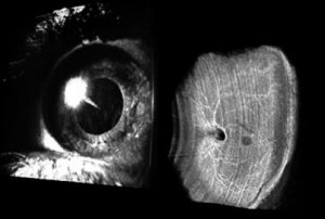
We developed a “Whole Eye” scanner that is based on Optical Coherence Tomography (OCT), which takes high-resolution, cross-sectional images of the eye. However, commercially available OCT systems currently only image the front or the back of the eye at a time, which limits our ability to accurately study global eye shape. The Whole-Eye OCT device addresses this issue by simultaneously imaging both, which allows us to obtain accurate measurements of subject eyes. The software we develop allows us to then use this information to study how the back of the eye changes with conditions such as near-sightedness and to improve surgical planning.
Watch our video on Whole-Eye imaging here.
Microscope-Integrated OCT
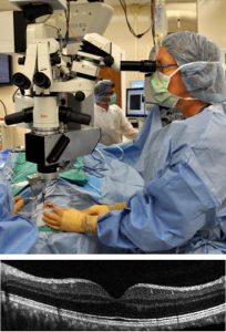
We are developing high-resolution, intraoperative ophthalmic imaging in collaboration with Drs. Cynthia Toth (Duke Eye Center) and Joseph Izatt (Duke Biomedical Engineering). Often times, ophthalmic surgeons only have a top-down view of the eye during operations. This makes it difficult to estimate how deep they are in the eye and can lead to surgical errors. With Microscope-Integrated OCT (MIOCT), physicians have live, cross-sectional visualization of the anatomic alterations they make during surgery. Not only does this allow the doctors to have better surgical orientation, but it also helps them detect pathologies during surgery.
Other
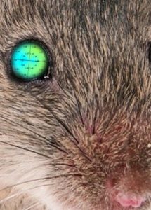
We have expanded our ophthalmic research to digitizing eyes from animal species as well. For instance, we developed OCT analysis software that created the first topography and thickness maps in mice, which will allow us to take better advantage of their research utility. This allows for improved characterization of genetically modified mice that replicate key features of human corneal diseases. Click here for the Mouse Corneal Analysis Program (MCAP).