
This summer, I am working in the Perfect Lab. The Perfect Lab focuses on understanding fungal pathogenesis at the molecular and genetic level. I am working specifically with the pathogen known as Cryptococcus neoformans which is a type of fungus that can infect different body sites, usually targeting the lungs or the central nervous system (consisting of the brain and spinal cord). An infection known as “cryptococcal meningitis” is developed when the fungus makes its way from the lungs to the brain. This fungus is the most common fungal infection that people with HIV/AIDS contract. Symptoms like headaches, fevers, neck pain, and more can arise once contracting cryptococcal meningitis.
My current project for the summer will consist of analyzing in a deeper context the cell capsule of Cryptococcus and seeing if the variation in capsule size contributes to the survival of the pathogen cell: does it benefit or weaken the cell to survive? The capsule plays a big part in the overall structure of the fungal cell because it serves as a protective layer against host phagocytic cells (a type of white blood cell that through phagocytosis engulfs bacteria, foreign particles, and dying cells to protect the body) and interferes with host immune mechanisms which can eliminate the pathogen from the body system, therefore the capsule helps the fungal cell to grow and develop further. (In some cases, the capsule can be so large that phagocytic cells can’t engulf the yeast cell).
Process of the experiment: To begin, I prepared the food (YPD plates) from scratch to grow C. neoformans isolates that originally came from patients in Southern Africa who had contracted cryptococcal meningitis. I learned the importance of keeping the YPD plates sterile and dry, so I grow this fungus and not an air contaminant that decided to become a food invader. So, yes space invaders do exist.
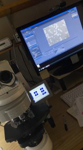
Once a batch of YPD plates had been made, they were kept on a table with a plastic cover on top to prevent any contamination. After letting the YPD plates sit for a few days, the next step to take was to streak C. neoformans Ambition isolates onto the YPD plates to grow the cell colonies of that strain onto the YPD plates. Once the plates were streaked, they are then placed inside an incubator at a temperature of 30 degrees Celsius to allow colony growth for 2–3 days. On Day 3 of growth, it is time to inoculate. The yeast cells grew well after just 3 days of growth. In this process, I transferred a single & isolated colony from the YPD plate into a tube filled with 3 mL of a SAB-MOPS media that has limited nutrients and is buffered to pH 7.3. After acquiring a colony using a sterile wooden applicator, I placed it onto the SAB-MOPS medium inside the tube to transfer the cells into the medium, then placed the tube into a vortex to mix the colony well into the media, and onto a shaker the tube went where it stayed for 5 days at a temperature of 30 degrees Celsius and 225 rpm (revolutions per minute). After the 5 days of growth on the shaker, the tube is removed. Next was staining the cells with India ink, which helps to detect the capsule that surrounds the yeast cell. A clear halo appears around the yeast cell that is the capsule. The cell suspension is transferred to microfuge tube and four microliters of India ink is added to 20 microliters of the cell suspension, as the last step. Four microliters were transferred onto a microscope slide, a cover slip was placed on top, and then taken onto the microscope for imaging.
With microscope imaging, the C. neoformans cells were made visible through magnification and their capsules as well. The initial images were not contrasted enough, so one change that I made was to increase the amount of India ink to 5 microliters. This increase in India ink made a big difference because the capsules were more visible under the microscope. For the analysis of the cells this helps a lot because when the sizes of the capsules, cell wall, and cell are being measured, a clear image outline of each capsule and cell will facilitate the measuring. It was so interesting to see how the strains varied in sizes and population growth. Some strains consisted of a tiny cell with a big capsule, while others had a medium-sized cell with not a lot of capsule surrounding it. The cell wall will also be looked at, thanks to the addition of a solution known as “Calcofluor White Stain” (CWS) which enables it to be seen under the microscope. CWS outlines the shape of the cell but not the capsule, and the variation in cell size may also affect virulence.
An additional observation that I made after imaging more strains was noticed that there the capsule size was not increasing or that there were some but not all cells producing capsule. There are different methods used to increase capsule production. I chose to evaluate the effect of temperature. The strains grown at a temperature of 30 degrees Celsius would be compared to strains grown at a temperature of 37 degrees to see if it would impact their cell or capsule growth. For this experiment, I’ll be using H99 which is a well-studied clinical strain of the pathogen, and later seeing the effect among with the Ambition isolates. If a certain temperature impacts cell growth and that of the capsule, it will be made an important factor to test our ultimate hypothesis of: Does variation in capsule size contribute to the level of survival of this pathogen?: does it benefit its survival? Temperature could be a contributing factor about whether a C. neoformans survives and continues to harm the body’s system as it can result in the increase of capsule resulting in a yeast cell beingmore resistant to the host defenses. I await the data that will come from the microscopic imaging and can’t wait to see what it reveals.
There are many factors that contribute to virulence and the survival of C. neoformans in the host. Another external factor that I’ll be evaluating with my research is seeing how its sensitivity to environmental stresses affects its growth. The two cell stressors that I will test are the sensitivity of C. neoformans isolates to caffeine and SDS (which is a type of detergent found in some soaps that makes the soap suds). The Ambition isolates of the pathogen C. neoformans will be put under the conditions of these two cell stressors, and I’ll monitor their growth over time. It will be interesting to compare isolates that are sensitive to SDS and caffeine for their capsule size. I can’t post any data as of now as I’m still collecting but in the coming weeks, I hope to gather data that can tell us more.
Very well done. It is clear you have a very good handle on your project. You did a really good job introducing the “problem”, then explaining how you are addressing this with your experiment and why your project is applicable. Great job!