Photo 1. Aaron waiting to give his dissertation presentation in the Bryan Research Building.
The Acharya Lab at Duke University is delighted to congratulate Dr. Aaron May on the successful defense of his doctoral dissertation, “Characterization of Novel and Diverse Henipavirus Glycoproteins,” presented to the Duke University Department of Biochemistry on October 28, 2025!
Aaron’s defense marks a special milestone as he becomes the first PhD graduate from the Acharya Lab. Aaron joined the Biochemistry PhD program in 2020 after completing his B.S. at the University of Wisconsin–Madison and became a member of the Acharya Lab in 2021. He was mentored by Dr. Priyamvada Acharya and personally thanked his committee members — Drs. Alberto Bartesaghi, Pei Zhou, Micah Luftig, and Kevin Saunders — for their support and guidance throughout his PhD journey.
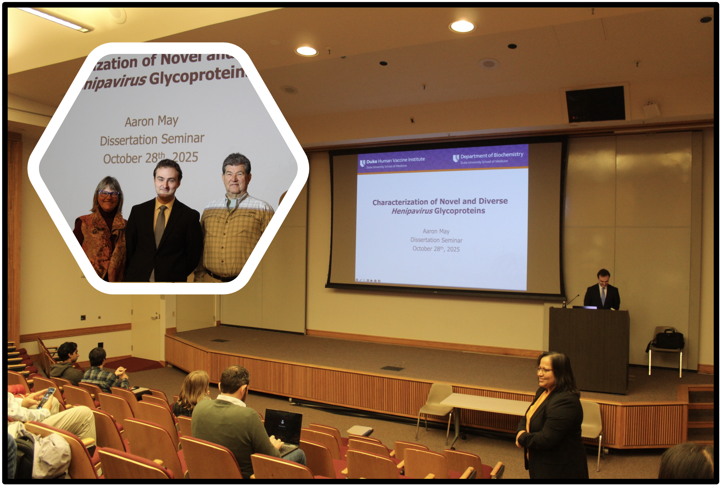
Photo 2. 11:55 AM at the 103 Bryan Research Building lecture hall—five minutes before the official start of Aaron’s dissertation. The hexagonal photo features Aaron with his parents.
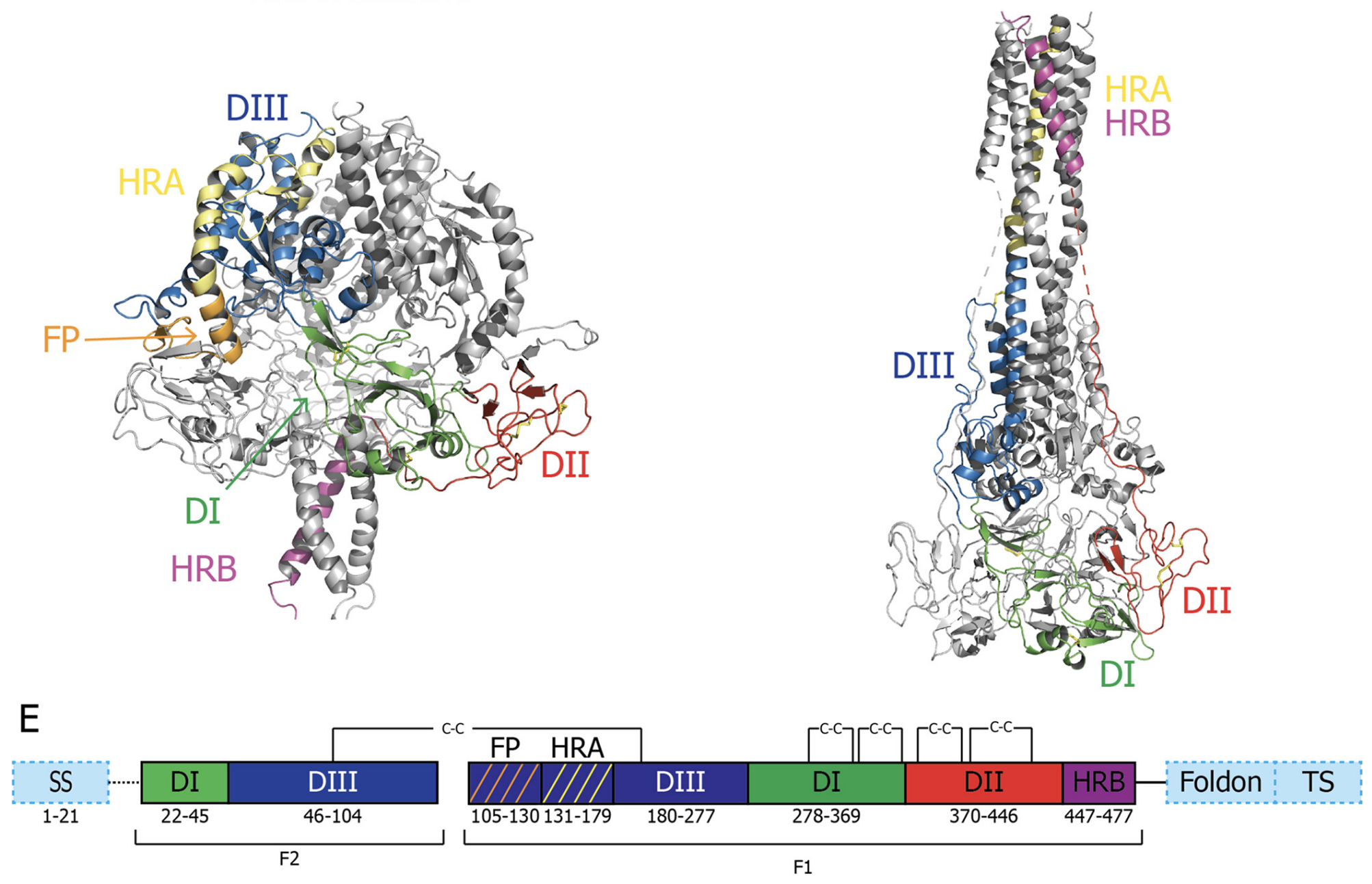
Fig 1. May AJ, et al. Structures of Langya Virus Fusion Protein Ectodomain in Pre- and Postfusion Conformation. J Virol. 2023
Among Aaron’s significant research contributions is his study titled “Structures of Langya Virus Fusion Protein Ectodomain in Pre- and Postfusion Conformation” (J. Virol., 2023), which revealed cryo-EM structures of the Langya virus fusion protein in both prefusion and postfusion conformations. This work identified unique surface features and fusion mechanisms that expand our understanding of Henipavirus evolution and inform future vaccine design. His most recent study, “Structural and Antigenic Characterization of Novel and Diverse Henipavirus Glycoproteins”, currently under review, builds on this foundation by examining a broad panel of henipavirus antigens.
Beyond his primary thesis project, Aaron also contributed to collaborative efforts in the lab, including structural studies on SARS-CoV-2, further demonstrating his versatility and dedication to advancing viral structural biology.

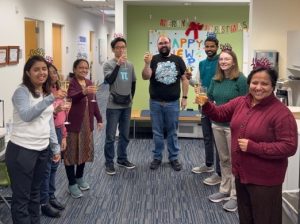
Photo 3. Celebrating Aaron’s successful defense. (Left) Dr. Priyamvada Acharya and Dr. Aaron J May. (Right) Aaron raising his glass in a toast.
Throughout his graduate career, Aaron has been recognized for his scientific rigor, collaborative spirit, and mentorship of junior trainees in the lab. The Acharya Lab celebrates his hard work, perseverance, and many accomplishments during his PhD. Way to go, Dr. May!