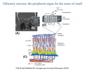
Schematic overview if the human olfactory mucosa. (A) coronal CT scan depicting location of the olfactory cleft. (B) zoomed image of olfactory cleft region. (C) Schematic of the olfactory neuroepithelium lining the olfactory cleft region, housing the olfactory sensory neurons and related cell populations. (D) Cilia projection from olfactory neurons into the nasal airspace contain olfactory receptors, G-protein couple receptors that are activated by binding of odor ligands.

Human olfactory biopsy analysis using single cell RNA-seq. Analysis of presbyosmic and normosmic patient biopsies identifies gene expression changes in olfactory basal stem cells, suggesting that inflammatory wear-and-tear may contribute to altered epithelial homeostasis in aging related olfactory loss.