Following the people and events that make up the research community at Duke

Students exploring the Innovation Co-Lab

Author: Olivia Zhu Page 2 of 3

The Science of Self-Agency: Dr. Nicolelis and the Walk Again Project

By Olivia Zhu

Screen grab from Univision of Juliano's robo-kick at the World Cup opening ceremony.

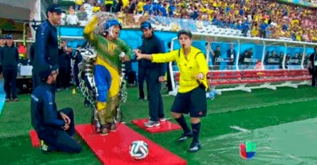
Screen grab from Univision of Juliano’s robo-kick at the World Cup opening ceremony.

Over the course of his 20-year career, Dr. Miguel Nicolelis has restored movement and self-agency to paraplegic patients.

On November 11th, as part of the Grand Challenge Seminar Series, Dr. Nicolelis captivated his audience by explaining the extensive process that culminated in Juliano, a Brazilian 29-year-old paralyzed from the chest downward in a car accident, performing the opening kick of the World Cup simply by using his mind.

Dr. Nicolelis has several faculty appointments in the Duke School of Medicine, Department of Psychology and Neuroscience, Institute for Brain Sciences, and Center for Neuroengineering. He has also written a book, Beyond Boundaries, about his work. His program, Walk Again, is supported by the Edmond and Lily Safia International Institute of Neuroscience in Brazil.

Dr. Nicolelis began making progress in 1999-2000 at Duke by developing electrodes that could record firing from multiple neurons. Using this technology, he determined which neurons were necessary for a monkey to move a joystick during a video game. Then, Dr. Nicolelis focused on creating a bypass that would bridge the mind directly to a computer, essentially removing the body as an intermediary.

He called this bypass a “Brain-Machine Interface,” or BMI, a term he coined at a cheese steak joint outside of Philadelphia. With the BMI, Dr. Nicolelis’s monkeys could play the video game without moving their arms or the joystick—they simply imagined themselves moving the joystick. The monkeys could even use their arms to do other tasks like eat or scratch themselves, creating a “third arm.”

Since then, with an extensive team of engineers, Dr. Nicolelis has implemented this technology by creating a IMG_1941hydraulically-powered exoskeleton that interprets a patient’s firing neurons and moves a patient’s legs accordingly.

He has also created artificial “skin,” which provides tactile feedback of movement to a patient’s upper body or, eventually, through an implant directly to the tactile cortex of the brain.

The technology is so accurate that patients report feeling “ghost limbs”—they believe that their legs are actually walking. The legendary Brazilian soccer player, Ronaldo, reportedly exclaimed “I’m moving!” with incredulity, when he was strapped to a chair testing Nicolelis’s technology.

Training with the exoskeleton also improves patients’ cardiovascular circulation, mental health, gastrointestinal health, and sensitivity in previously paralyzed areas.

Dr. Nicolelis is truly using science to stretch the boundaries of the human body.

Artistic Anatomy: An Exploration of the Spine

By Olivia Zhu

How many times have you acted out the shape of a vertebra with your body? How many times have you even imagined what each of your vertebrae looks like?

On Wednesday, October 1, Kate Trammell and Sharon Babcock held a workshop on the spine as part of the series, Namely Muscles. In the interactive session, they pushed their audience members to gain a greater awareness of their spines.

Participants assemble vertebrae and discs of the spine

Participants assemble vertebrae and discs of the spine

Trammell and Babcock aim to revolutionize the teaching of anatomy by combining art, mainly through dance, and science. They imagine that a more active, participatory learning style will allow students from all backgrounds to learn and retain anatomy information much better. Babcock, who received her Ph.D. in anatomy from Duke, emphasized how her collaboration with Trammell, a dancer and choreographer, allowed her to truly internalize her study of anatomy. The workshop participants, who included dancers and scientists alike, also reflected a fusion of art and science.

Trammell observes the living sculptures of thoracic vertebrae

Trammell observes the living sculptures of thoracic vertebrae

To begin the exploration of the spine, Trammell and Babcock had participants close their eyes and feel models of individual vertebrae to gain tactile perception. Trammell and Babcock then instructed participants to make the shape of the vertebrae they felt with their bodies, creating a living sculpture garden of various interpretations of vertebrae–they pointed out key aspects of vertebrae as they walked through the sculptures.

Finally, Trammell and Babcock taught movement: in small groups, people played the roles of muscles, vertebrae, and spinal discs. They worked on interacting with accurate movements (for example, muscles only pull; they cannot push) to illustrate different movements of the spine.

Interactive illustration of a muscle pulling vertebrae

Interactive illustration of a muscle pulling vertebrae


To complete the series, Trammell performed Namely, Muscles, choreographed by Claire Porter, on October 4th  at the Ark.

An Intersection of Math and Medicine: Modeling Cancerous Tumor Kinetics

Anne Talkington with the MAMS function

Anne Talkington with the MAMS function

By Olivia Zhu

Anne Talkington, an undergraduate Mathematics student under the auspices of Richard Durrett, attempts to gain a quantitative grasp on cancer through mathematical modeling. Historically, tumor growth has only been measured in vitro (in a laboratory setting); however, Talkington looks at clinical data from MRIs and mammograms to study how tumors grow in vivo (in the human body).

Talkington is primarily interested in how fast tumors grow and if growth is limited. To analyze these trends, Talkington extracted two time-point measurements of tumor size — one at diagnosis and one immediately before treatment — and compared their change to a variety of mathematical functions. She studied unlimited functions, including the exponential, the power law, and the 2/3 power law, which represents growth limited by surface area, as well as limited functions, including the generalized logistic, which has an upper growth limit, and the Gompertz. Her favorite function is an unlimited function that she created called the Modified Alternating Maclaurin Series, or MAMS, which she originally intended to model microbial growth.

Talkington also examined various types of cancer: breast cancer, liver cancer, tumors of the nerve that connect the ear to the brain, and meningioma, or tumors of the membranes that surround the brain and spinal cord. She expected growth rates among clinical groups to be constant, but she did not generalize between the groups due to demographic bias and other confounding factors.

Ultimately, Talkington found that breast cancer and liver cancer grew exponentially, while tumors of the meninges or vestibulocochlear nerve grew according to the 2/3 power law. Talkington’s work in model-fitting cancer growth will facilitate the administration of effective treatment, which is often growth-stage dependent.

The Mystery Behind the Camel Statue

Knut Schmidt-Nielsen

A file photo of the real Knut Schmidt-Nielsen, not the bronze one, standing with the enigmatic camel statue dedicated to him and his work.

By Olivia Zhu           

The camel statue between the Biology Building and Gross Hall is a staple of Duke’s campus, but the significance behind this landmark is generally unknown.

On Monday, September 22, faculty from the Biology Department gathered for a dedication to remember the man behind the camel statue (or rather, in front of it), Dr. Knut Schmidt-Nielsen, who died in 2007.

Knut Schmidt-Nielsen, who would have turned 99 this Wednesday, was “the father of comparative physiology and integrative biology” and a James B. Duke professor at Duke’s Biology Department starting in 1952.

Schmidt-Nielsen studied the physiology of the camel’s nose, received the International Prize for Biology, and wrote the authoritative text on animal physiology.

Dr. Stephen Wainwright, who was present at the dedication, commissioned the camel to British sculptor Jonathan Kingdon, who finished the bronze camel statue in 1993. The inscription for the statue, “Tell me about yourself, Camel, that I may know myself,” encapsulates Schmidt-Nielsen’s outlook on physiology.

According to Dr. Steven Vogel, who was recruited to Duke’s faculty by Schmidt-Nielsen 49 years ago, Schmidt-Nielsen was actually shy and rather uncomfortable with the statue of himself. Vogel reported that Schmidt-Nielsen greatly advanced the zoology department with his high standards and “great charm and urbanity.”

“You could never say no to Knut,” Vogel said. Schmidt-Nielsen was also reportedly  “a very serious wine drinker”—accordingly, the dedication ceremony ended with wine and champagne.

To learn more about Knut Schmidt-Nielsen, read Vogel’s memoirs or a recommended autobiography, The Camel’s Nose.

Knut Schmidt-Nielsen

The statue as it appears now, with Knut in bronze. (File photo)

Mathematical Restoration of Renaissance Masterpieces

Screen Shot 2014-09-21 at 10.41.22 PM

The Ghissi Masterpiece, missing the ninth panel

By Olivia Zhu

Ninth panel of the Ghissi masterpiece, as reconstructed by Charlotte Caspers

Ninth panel of the Ghissi masterpiece, as reconstructed by Charlotte Caspers

What do Renaissance masterpieces and modern medical images have in common?

The same mathematical technique, “oriented elongated filters,” originally developed to detect blood vessels in medical images can actually be used to detect cracks in digital images of antiquated Renaissance paintings.

On September 19, Henry Yan, Rowena Gan, and Ethan Levine, three undergraduate students at Duke, presented their work on oriented elongated filters and many other techniques to the Math Department. Yan, Gan, and Levine performed summer research to detect and correct cracks in the digitized Ghissi masterpiece, an altarpiece done by 14-century Italian painter Francescuccio di Cecco Ghissi. The altarpiece originally consisted of nine panels, but one was lost in the annals of history and has been recently reconstructed by artist and art historian Charlotte Caspers.

The role of the three undergrads was to digitally rejuvenate the panels of the Ghissi masterpiece, which had faded and accumulated cracks in paint layers because of weathering factors like pressure and temperature. Using various mathematical analysis techniques based in Matlab, including oriented elongated filters, linear combinations of 2-D

Henry Yan's K-SVD analysis to detect cracks in the image at left

Henry Yan’s K-SVD analysis to detect cracks in the image at left

Gaussian kernels (which essentially create directional filters), K-SVD (which updates atoms to better fit an image), and multi-scale top-hat (which extracts small elements and details from an image), the research group created a “crack map,” which they overlaid on the original image.

Then they instructed the computer to fill in the cracks with the colors directly adjacent to the cracks, thereby creating a smoother, crack-free image—this method is called inpainting.

In the future, Yan, Gan, and Levine hope to optimize the procedures they have already developed to accomplish color remapping to digitally age or refurbish images so that they look contemporary to their historical period, and to digitally restore gilding, the presence of gold leaf on paintings.

A Quiet but Fundamental Case in the Flame Retardant Debate

By Olivia ZhuClose up of sofa in the living room

The dangers of flame retardants have long been the source of public health debate, thrust into the public eye by legislators, non-profit organizations, and special interest groups.

Often missing, though, is the story of the extensive scientific background necessary to ground these arguments: Heather Stapleton, the Dan and Bunny Gabel Associate Professor of Environmental Ethics and Sustainable Environmental Management, and research scientist Ellen Cooper and their teams  fill this role at the Nicholas School of the Environment.

As a former intern at the Center for Environmental Health, where I advocated against legislation that promotes the use of flame retardant chemicals, I was fascinated to learn the technical side of the conversation from Cooper.

Flame retardants have been present in furniture and other foam-based items largely due to fire safety regulations like California’s TB117, which requires home furniture to resist bursting into flame for a certain amount of time. Historically, manufacturers have met this standard by using flame retardant chemicals that may impact brain development in children or even cause cancer. Consequently, foam producers have added flame retardants to all their foam, even foam meant for products not covered under TB117. Evidently, the resulting flame retardant-laden selection of furniture poses a threat to the health of consumers, who effectively have little choice in protecting themselves against potentially dangerous chemicals.

Nicholas school environmental chemist Heather Stapleton in the lab. (Duke Photography)

Nicholas school environmental chemist Heather Stapleton in the lab. (Duke Photography)

Unsurprisingly, scientifically studying flame retardants requires rigorous chemical analysis. Ellen Cooper, along with the Analytical Chemistry Core, specializes in preparing foam samples and running them through mass spectrometers to identify the flame retardants. The Chemistry Core collaborates with the Stapleton Lab to ensure accurate analysis of often-fragile flame retardants. Both are a part of Duke’s Superfund Research Center.

Cooper’s work has been instrumental in the Stapleton lab’s creation of a foam testing service for consumers. In January, the Stapleton lab started the first foam testing service ever open to the public! Now, consumers can send in up to five samples from their furniture to receive complimentary results which are provided by the Analytical Chemistry Core. On the foam testing website, consumers can also find statistics on common sources of flame retardants and information on how to avoid exposure to flame retardants. Through these efforts, the Stapleton lab is allowing consumers to take back their autonomy.

Additionally, Cooper is working to create a database of all collected samples. Currently, there exists no definite data on which manufacturers’ products contain which flame retardants, or how levels of flame retardants in furniture have changed over time. With enough data points,  Cooper and her team hope to create such heat maps or time analyses, making for a more informed debate, and ultimately a more well-protected public.

Summer Restoration in a Bolivian Winter

By Olivia Zhu2014-06-19 10.45.15

My biggest accomplishment this summer was being able to call the mountains of Bolivia home. Far away from the lecture halls of Duke, I encountered a profound, alternative education that included everything from learning traditional dances to working in a rural hospital laboratory to raising pigs.

Of course, living in Bolivia for two months had its challenges, like a diet in which potatoes were considered vegetables, repeated food poisoning from chicha, the local alcoholic drink consisting of fermented corn, lack of a consistent water source, many near-car accidents, and most of all a deep-seated machismo, but I feel that these were all almost inextricable aspects of a culture that left such a positive impression upon me.

El Hospital Pietro Gamba in Anzaldo, Bolivia

El Hospital Pietro Gamba in Anzaldo served over 69 rural communities in Bolivia

Of course, the inextricability of such factors posed a problem for me as an intern at El Hospital Pietro Gamba encouraging sustainable development to promote public health. Although 80% of children had head lice, a vast majority contracted repeated gastrointestinal bacterial infections, and countless had scabies, the community seemed to get along contentedly. Regardless, with support from the Foundation for Sustainable Development and DukeEngage, my sponsor organizations, I leveraged the relatively new running water system, implemented only 25 years ago, to set in motion a comprehensive lice campaign, to obtain government funding of soap in public restrooms for at least two years, and to create preventative medicine informational materials.

The majority of my education, though, occurred outside the scope of my project. Most importantly, I’ve learned to openly embrace different forms of learning, like relaxation or soccer, that energize me to wholeheartedly pursue my rigorous biophysics career, which I am so fortunate to have at one of the best universities in the world.

The idea of the Aymara New Year illustrates my mentality poignantly: on the first day of the Aymara New Year, traditional Bolivians wish for health, prosperity, and happiness, just as we do in the United States. However, they have a deeper connection with Pachamama, or Mother Nature: on New Year’s Day, they wake up early in the morning to stand on the ground barefoot, awaiting the first rays of the sun. They believe that watching these rays rise above the horizon and light the earth will bring them energy for the entire year. In this, the Aymara New Year represents both personal aspiration and attenuation with the environment.

Similarly, I now aim to maintain a balance between self and surroundings: I hope to be more attuned to the world around me rather than single-mindedly submersing myself in quantum physics, as I believe that varied experiences will infuse me with energy in whatever I pursue. Now, back at Duke for the start of my junior year, I’m excited to begin blogging again and to continue my adventures and education here on campus.

The Aymara sunrise on June 21, 2014.

The Aymara sunrise on June 21, 2014.

Studying patterns in bacterial organization

credit to Gerard Wong, of the California NanoSystems Institute

credit to Gerard Wong, of the California NanoSystems Institute

by Olivia Zhu

Bacterial biofilms, at first glance, may seem to be spontaneous, random phenomena from which we have no power to protect our environment or ourselves.

They’re potentially useful as an aid to wastewater treatment, but they also cause infections that account for $6 billion a year in health care costs. Biofilms are also more resistant to antibiotic drugs, making them difficult to eradicate.

Dr. Kun Zhao, of the California NanoSystems Institute at UCLA, refuses to see biofilms as arbitrary: he emphasizes the fact that biofilms are communities of bacteria in self-produced polymeric matrices of polysaccharides, and using a biophysical approach, he studies the pattern behind their organization.

Central questions in Zhao’s research include how bacterial colonies transition from reversible to irreversible attachment, how they migrate, and how they ultimately disperse. Specifically, Zhao examines the polysaccharide Psl, which poses a positive feedback loop because it is both secreted by moving bacteria and serves as a chemo-attractant for future bacteria movement. The positive feedback creates an inherent pattern, as bacteria are more likely to visit a location they have been to before.

Zhao and colleagues have also discovered that bacterial mutants that cannot produce Psl exhibit more random and uniform movement.

To better quantify bacterial movement,Zhao has created a computer algorithm that shows the full movement history of each individual bacterium on a dish, and that provides a “search engine” allowing researchers to find every bacterium performing specific life cycle activities, like division.

Zhao has postulated a “rich get richer” mechanism for biofilms. He compares bacterial organization to Wall Street because concentrated movement ensures that some cells become extremely enriched. In the future, he hopes to model colloidal structures for biological problems, like the growth of the bacterial cell wall. Zhao currently uses colloids, which in physics are used as models for atomic systems, to observe how shapes affect self-assembly. He also would like to look at cell-substrate interactions, which are implicated in bacterial territoriality and social interactions.

Discovering “CRISPR” methods for genetic recombination

Screen Shot 2014-03-27 at 9.54.19 PM

By Olivia Zhu

In a lecture to an overflowing auditorium in the Bryan Research Building on March 27th, Dr. Jennifer Doudna, of the University of California, Berkeley, unraveled her story of research into CRISPRs, or “clustered regularly interspaced short palindromic repeats.” Dr. Doudna specializes in RNA; she started her project on CRISPRs seven years ago, when CRISPRs were denounced as no more than junk.

The CRISPR method includes a modifiable RNA sequence whose function is to recognize target sequences on DNA. The RNA also includes a target sequence that induces cleavage by the associated protein, CAS9. CAS9 introduces double-stranded breaks and represents an exciting improvement over the previous, less efficient collection of nine proteins used to cleave DNA; the breaks make room for insertion of new genes. The CRISPR-CAS9 system has inserted genes into a wide range of organisms, including bacteria, yeast, nematode worms, fruit flies, plants, fish, mice, and even human cells.

Jennifer Doudna

Jennifer Doudna of UC Berkeley and the Howard Hughes Medical Institute

While researchers are actively investigating the possibility of using CRISPR technology to alter genes, Doudna said the mechanism behind CRISPR gene editing remains unclear. For example, it seems extraordinary that the CRISPR-CAS9 system can locate and unwind specific DNA sequences in human cells, as the DNA there is highly condensed around histones and methylated.

Doudna’s lab is working to understand the details of the CRISPR process. One current hypothesis includes the idea that there is a spring mechanism that allows the CAS9 protein to effectively cleave DNA strands.

Nevertheless, CRISPR technology has been instrumental in allowing more precise and efficient genetic modification. What we once considered junk has spurred substantial advances across various fields of science.

Rhyne King: Unearthing an Ancient Religion

rhyne

By Olivia Zhu

Rhyne King, a senior from Greensboro, North Carolina, plunges into the depths of history to retrieve remnants of long-past civilizations.  Rhyne is currently writing his senior thesis on the religion of the Achaemenid Empire of Persia, which existed in modern day Iran until 330 B.C.E., when Alexander the Great’s army conquered it.  Specifically, Rhyne is focusing on what the Achaemenid religion was and how the Achaemenids treated foreign religions.  Rhyne says that it is rather difficult to directly compare the Achaemenid religion to any modern religion, but that some imagine it to be similar to Zoroastrianism.

Rhyne’s skill set and dedication to his research are extraordinary in and of themselves.  After spending the summer studying with Professor Jacques Bromberg, Rhyne added the skill of reading Old Persian to his repertoire of languages, which includes Latin, Greek, and Persian.  He uses Old Persian, the language of the Achaemenid kings, to read inscriptions about their religion.  Rhyne has also traveled to the British Museum in London to inspect Achaemenid inscriptions and art.

An inscription Rhyne studied at the British Museum

An inscription Rhyne studied at the British Museum

Rhyne pioneers investigation into the Achaemenid empire by balancing Greek accounts, which currently form a majority of the body of knowledge about the civilization, with Persian sources.  He says that up to this point, historians have not reconciled the two sources systematically.  Rhyne is particularly fascinated by the Achaemenid empire’s tolerance of religion.  He emphasizes, however, that their tolerance, immortalized in the Cyrus inscription from Babylon, was not an ancient declaration of human rights; rather, it was the system that proved most convenient for them.

Throughout his four years at Duke, Rhyne has also served as president of the Round Table selective living group and the Latin Club, and he has played for the marching band.  Rhyne plans to continue researching the Achaemenid Empire in graduate school, and to someday become a professor.

Page 2 of 3

Powered by WordPress & Theme by Anders Norén