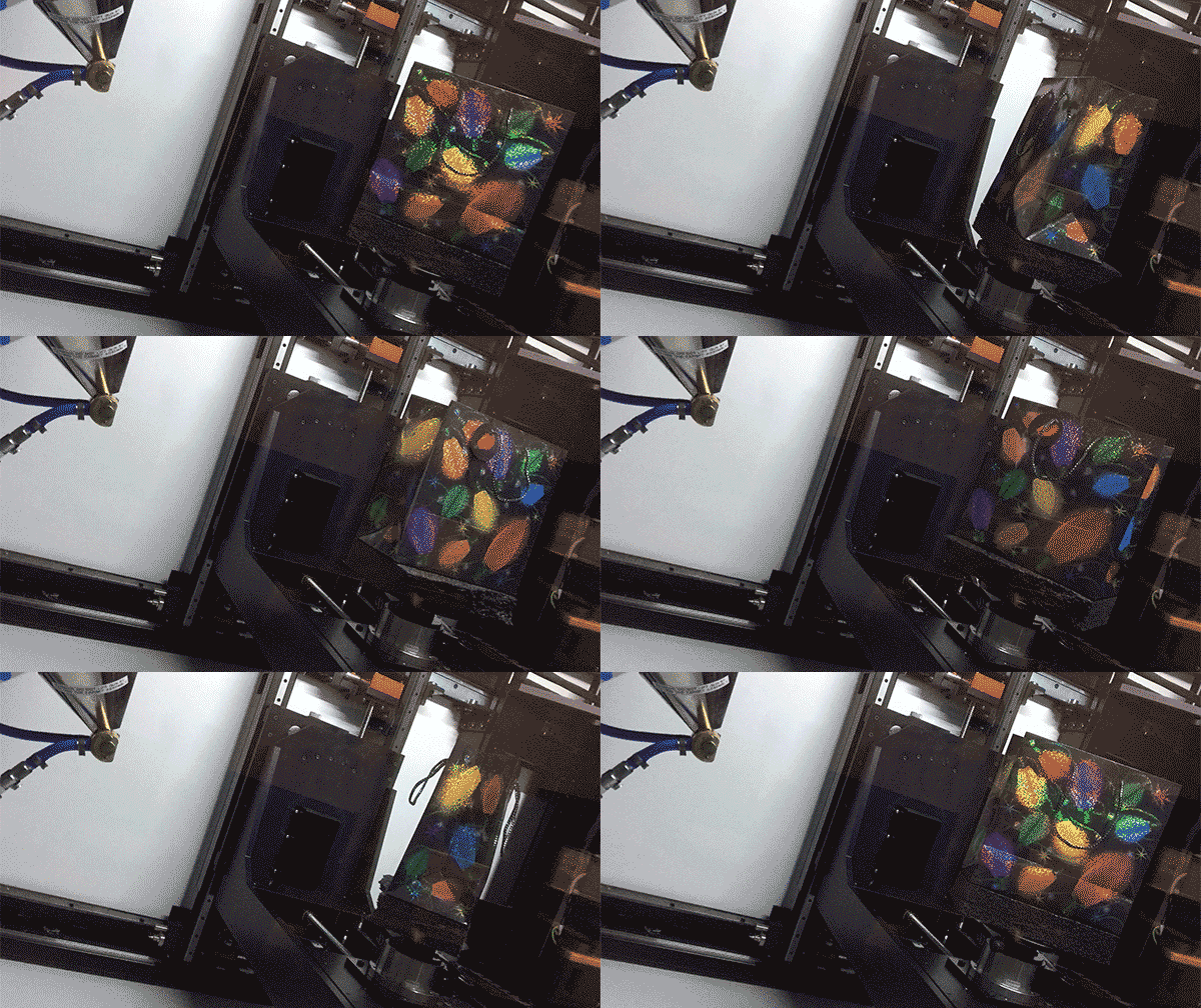
If, like me, you just cannot wait until Christmas morning to find out what goodies are hiding in those shiny packages under the tree, we have just the solution for you: stick them in a MicroCT scanner.

Our glittery package gets the X-ray treatment inside Duke’s MicroCT scanner. Credit Justin Gladman.
Micro computed-tomography (CT) scanners use X-ray beams and sophisticated visual reconstruction software to “see” into objects and create 3D images of their insides. In recent years, Duke’s MicroCT has been used to tackle some fascinating research projects, including digitizing fossils, reconstructing towers made of stars, peaking inside of 3D-printed electronic devices, and creating a gorgeous 3D reconstruction of organs and muscle tissue inside this Southeast Asian Tree Shrew.

A 20 minute scan revealed a devilish-looking rubber duck. Credit Justin Gladman.
But when engineer Justin Gladman offered to give us a demo of the machine last week, we both agreed there was only one object we wanted a glimpse inside: a sparkly holiday gift bag.
While securing the gift atop a small, rotating pedestal inside the device, Gladman explained how the device works. Like the big CT scanners you may have encountered at a hospital or clinic, the MicroCT uses X-rays to create a picture of the density of an object at different locations. By taking a series of these scans at different angles, a computer algorithm can then reconstruct a full 3D model of the density, revealing bones inside of animals, individual circuits inside electronics – or a present inside a box.
“Our machine is built to handle a lot of different specimens, from bees to mechanical parts to computer chips, so we have a little bit of a jack-of-all-trades,” Gladman said.
Within a few moments of sticking the package in the beam, a 2D image of the object in the bag appears on the screen. It looks kind of like the Stay Puft Marshmallow Man, but wait – are those horns?

Blue devil ducky in the flesh.
Gladman sets up a full 3D scan of the gift package, and after 20 minutes, the contents of our holiday loot is clear. We have a blue devil rubber ducky on our hands!
Blue ducky is a fun example, but the SMIF lab always welcomes new users, Gladman says, especially students and researchers with creative new applications for the equipment. For more information on how to use Duke’s MicroCT, contact Justin Gladman or visit the Duke SMIF lab at their website, Facebook, Youtube or Instagram pages.

Post by Kara Manke
