Following the people and events that make up the research community at Duke

Students exploring the Innovation Co-Lab

Month: December 2016

Life Lessons from a Neuroscientist

I recently had the privilege of sitting down with Dr. Anne Buckley, a professor and  neuropathologist working in Dr. Chay Kuo’s cell biology lab at Duke. I got a first-hand account of her research on neuron development and function in mice. But just as fascinating to me were the life lessons she had learned during her time as a researcher.

Anne Buckley, M.D. Ph.D., is an assistant professor of pathology

Anne Buckley, M.D. Ph.D., is an assistant professor of pathology

Buckley’s research looks at brain tumors in mice. She recently found that some of the mice developed the tumors in an area full of neurons, the roof of the fourth ventricle, which is of particular interest because humans have developed tumors in the same location. This discovery could show how neurological pathways affect tumor formation and progression.

Buckley also gave me some critical words of advice, cautioning me that research isn’t for everyone.

“Research is not glamorous, and not always rewarding,” she warned me. When she first started research, Buckley learned a hard lesson: work doesn’t necessarily lead to results. “For every question I went after, I found ten more unresolved,” she said. “To be a researcher, it takes a lot of perseverance and resilience. A lot of long nights.”

But that’s also the beauty of research. Buckley says that she’s learned to find happiness in the small successes, and that she “enjoys the process, enjoys the challenge.”

And when discoveries happen?

“When I look at data, and I see something unexpected, I get really excited,” she says. “I know something that no one else knows. Tomorrow, everyone will know. But tonight, I’m the only person in the world who knows.”

kendra_zhong_headshotGuest Post by Kendra Zhong, North Carolina School of Science and Math, Class of 2017

Evolutionary Genetics Shaping Health and Behavior

Dr. Jenny Tung is interested in the connections between genes and behavior: How does behavior influence genetic variation and regulation and how do genetic differences influence behavior?

A young Amboseli baboon hitches a ride with its mother. (Photo by Noah Snyder-Mackler)

A young Amboseli baboon hitches a ride with its mother. (Photo by Noah Snyder-Mackler)

An assistant professor in the Departments of Evolutionary Anthropology and Biology at Duke, Tung is interested in evolution because it gives us a window into why the living world is the way it is. It explains how organisms relate to one another and their environment. Genetics explains the actual molecular foundation for evolutionary change, and it gives part of the answer for trait variation. Tung was drawn as an undergrad towards the combination of evolution and genetics to explain every living thing we see around us; she loves the explanatory power and elegance to it.

Tung’s longest collaborative project is the Amboseli Baboon Research Project (ABRP), located in the Amboseli ecosystem of East Africa. She co-leads it with Susan Alberts, chair of evolutionary anthropology at Duke, Jeanne Altmann at Princeton, and Beth Archie at Notre Dame.

Tung has spent months at a time on the savannah next to Mount Kilimanjaro for this project. The ABRP monitors hundreds of baboons in several social groups and studies social processes at several levels. Recently the project has begun to include genetics and other aspects of baboon biology, including the social behaviors within the social groups and populations, and how these behaviors have changed along with the changing Amboseli ecosystem. Tung enjoys different aspects of all of her projects, but is incredibly grateful to be a part of the long-term Amboseli study.

Jenny Tung

Jenny Tung is an assistant professor in evolutionary anthropology and biology.

The process of discovery excites Tung. It is hard for her to pin down a single thing that makes research worth it, but “new analyses, discussions with students who teach me something new, seeing a great talk that makes you think in a different way or gives you new research directions to pursue” are all very exciting, she said.

Depending on the project, the fun part varies for her; watching a student develop as a scientist through their own project is rewarding, and she loves collaborating with extraordinary scientists. Specific sets of collaborators make the research worth it. “When collaborations work, you really push each other to be better scientists and researchers,” Tung said.

Raechel ZellerGuest post by Raechel Zeller, North Carolina School of Science and Math, Class of 2017

X-mas Under X-ray

If, like me, you just cannot wait until Christmas morning to find out what goodies are hiding in those shiny packages under the tree, we have just the solution for you: stick them in a MicroCT scanner.

A christmas present inside a MicroCT scanner.

Our glittery package gets the X-ray treatment inside Duke’s MicroCT scanner. Credit Justin Gladman.

Micro computed-tomography (CT) scanners use X-ray beams and sophisticated visual reconstruction software to “see” into objects and create 3D images of their insides. In recent years, Duke’s MicroCT has been used to tackle some fascinating research projects, including digitizing fossils, reconstructing towers made of stars, peaking inside of 3D-printed electronic devices, and creating a gorgeous 3D reconstruction of organs and muscle tissue inside this Southeast Asian Tree Shrew.

x-ray-view

A 20 minute scan revealed a devilish-looking rubber duck. Credit Justin Gladman.

But when engineer Justin Gladman offered to give us a demo of the machine last week, we both agreed there was only one object we wanted a glimpse inside: a sparkly holiday gift bag.

While securing the gift atop a small, rotating pedestal inside the device, Gladman explained how the device works. Like the big CT scanners you may have encountered at a hospital or clinic, the MicroCT uses X-rays to create a picture of the density of an object at different locations. By taking a series of these scans at different angles, a computer algorithm can then reconstruct a full 3D model of the density, revealing bones inside of animals, individual circuits inside electronics – or a present inside a box.

“Our machine is built to handle a lot of different specimens, from bees to mechanical parts to computer chips, so we have a little bit of a jack-of-all-trades,” Gladman said.

Within a few moments of sticking the package in the beam, a 2D image of the object in the bag appears on the screen. It looks kind of like the Stay Puft Marshmallow Man, but wait – are those horns?

Blue devil ducky in the flesh.

Blue devil ducky in the flesh.

Gladman sets up a full 3D scan of the gift package, and after 20 minutes, the contents of our holiday loot is clear. We have a blue devil rubber ducky on our hands!

Blue ducky is a fun example, but the SMIF lab always welcomes new users, Gladman says, especially students and researchers with creative new applications for the equipment. For more information on how to use Duke’s MicroCT, contact Justin Gladman or visit the Duke SMIF lab at their website, Facebook, Youtube or Instagram pages.

Kara J. Manke, PhD

Post by Kara Manke

When Art Tackles the Invisibly Small

Huddled in a small cinderblock room in the basement of Hudson Hall, visual artist Raewyn Turner and mechatronics engineer Brian Harris watch as Duke postdoc Nick Geitner positions a glass slide under the bulky eyepiece of an optical microscope.

To the naked eye, the slide is completely clean. But after some careful adjustments of the microscope, a field of technicolor spots splashes across the viewfinder. Each point shows light scattering off one of the thousands of silver nanoparticles spread in a thin sheet across the glass.

“It’s beautiful!” Turner said. “They look like a starry sky.”

AgAlgae_40x_Enhanced3

A field of 10-nanometer diameter silver nanoparticles (blue points) and clusters of 2-4 nanoparticles (other colored points) viewed under a dark-field hyperspectral microscope. The clear orbs are cells of live chlorella vulgaris algae. Image courtesy Nick Geitner.

Turner and Harris, New Zealand natives, have traveled halfway across the globe to meet with researchers at the Center for the Environmental Implications of Nanotechnology (CEINT). Here, they are learning all they can about nanoparticles: how scientists go about detecting these unimaginably small objects, and how these tiny bits of matter interact with humans, with the environment and with each other.

img_2842

The mesocosms, tucked deep in the Duke Forest, currently lay dormant.

The team hopes the insights they gather will inform the next phases of Steep, an ongoing project with science communicator Maryse de la Giroday which uses visual imagery to explore how humans interact with and “sense” the nanoparticles that are increasingly being used in our electronics, food, medicines, and even clothing.

“The general public, including ourselves, we don’t know anything about nanoparticles. We don’t understand them, we don’t know how to sense them, we don’t know where they are,” Turner said. “What we are trying to do is see how scientists sense nanoparticles, how they take data about them and translate it into sensory data.”

Duke Professor and CEINT member Mark Wiesner, who is Geitner’s postdoctoral advisor, serves as a scientific advisor on the project.

“Imagery is a challenge when talking about something that is too small to see,” Wiesner said. “Our mesocosm work provides an opportunity to visualize how were are investigating the interactions of nanomaterials with living systems, and our microscopy work provides some useful, if not beautiful images. But Raewyn has been brilliant in finding metaphors, cultural references, and accompanying images to get points across.”

img_2872

Graduate student Amalia Turner describes how she uses the dark-field microscope to characterize gold nanoparticles in soil. From left: Amalia Turner, Nick Geitner, Raewyn Turner, and Brian Harris.

On Tuesday, Geitner led the pair on a soggy tour of the mesocosms, 30 miniature coastal ecosystems tucked into the Duke Forest where researchers are finding out where nanoparticles go when released into the environment. After that, the group retreated to the relative warmth of the laboratory to peek at the particles under a microscope.

Even at 400 times magnification, the silver nanoparticles on the slide can’t really be “seen” in any detail, Geitner explained.

“It is sort of like looking at the stars,” Geitner said. “You can’t tell what is a big star and what is a small star because they are so far away, you just get that point of light.”

But the image still contains loads of information, Geitner added, because each particle scatters a different color of light depending on its size and shape: particles on their own shine a cool blue, while particles that have joined together in clusters appear green, orange or red.

During the week, Harris and Turner saw a number of other techniques for studying nanoparticles, including scanning electron microscopes and molecular dynamics simulations.

steepwashing-cake-copy-23

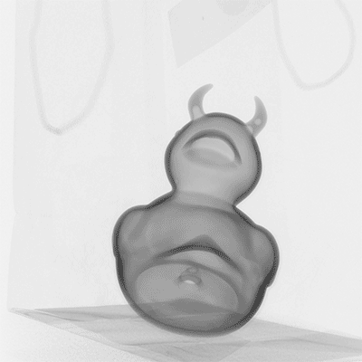
An image from the Steep collection, which uses visual imagery to explore how humans interact with the increasingly abundant gold nanoparticles in our environment. Credit: Raewyn Turner and Brian Harris.

“What we have found really, really interesting is that the nanoparticles have different properties,” Turner said. “Each type of nanoparticle is different to each other one, and it also depends on which environment you put them into, just like how a human will behave in different environments in different ways.”

Geitner says the experience has been illuminating for him, too. “I have never in my life thought of nanoparticles from this perspective before,” Geitner said. “A lot of their questions are about really, what is the difference when you get down to atoms, molecules, nanoparticles? They are all really, really small, but what does small mean?”

Kara J. Manke, PhD

Post by Kara Manke

José Jerónimo – Innovations in Cervical Cancer Screening

José Jerónimo and his team are transforming the face of cervical cancer screening. Jerónimo is a physician and senior advisor for the women’s cancers branch of PATH, an international nonprofit organization that uses innovative technologies to improve health outcomes in developing countries. Jerónimo, who’s work at PATH has facilitated the prevention and treatment of cervical cancer for thousands in the developing world, spoke at the Duke Institute for Global Health on Dec. 2.

Cervical cancer testing has been a point of conflict in the medical community for quite some time now, for the pap smear — for many years, the only test available to detect cervical cancer — is not very sensitive to abnormal tissue. Since skepticism with the pap smear arose a few decades ago, doctors like Jerónimo have been working tirelessly to find more effective screening strategies.

José Gerónimo, Peruvian physician and public health advocate, received his specialty training in gynecologic oncology at the National Cancer Institute in Peru.

José Jerónimo, Peruvian physician and public health advocate, received his specialty training in gynecologic oncology at the National Cancer Institute in Peru.

Cervical cancer can be acquired through the presence of HPV (human papilloma virus). Chronic infections of HPV have been proven to increase the likelihood of contracting cervical cancer, so developing primary prevention initiatives to avoid developing HPV to begin with are essential to decrease the prevalence of cervical cancer. HPV testing, unlike the pap smear, can be self-collected and does not require the complex, expensive machinery that the pap smear does. Initial self-sampling studies in India, Uganda, and Nicaragua indicated a willingness by the female community to self-test, so long as sanitary and private conditions were provided.

Studies in the Jujuy province of Argentina indicated that community health workers played a key role in facilitating the self-sampling process. When the health workers differed locals to clinics or sent them to facilities for testing, only 20 percent actually went. But, when they brought the self-sampling tests to locals’ homes directly, testing was above 80 percent. The easy accessibility of self-sampling, along with encouragement by local health volunteers, clearly showed that self-sampling was much more effective.

A group of female community health workers in Lima, Peru, educating the community about HPV testing.

Jerónimo’s current work focuses on strengthening government screening systems for HPV that are already in place. By helping ministries introduce and scale up the testing, he and others at PATH hope to decrease HPV and cervical cancer rates.

But, it goes beyond testing. Jerónimo emphasizes the need for evaluation and follow-up mechanisms after testing positive. Although testing efforts have improved significantly, the treatment provided after for those who have tested positive is still lagging. Jerónimo claims that much of this is due to minimal efforts by the local governments to really follow through beyond the testing phase.

PATH is looking for innovative ways to treat HPV that are inexpensive and effective. They recently developed their own version of the thermal coagulator, a probe that treats infected tissue using heat. Their design runs on a battery, rather than needing constant electricity, and uses a progressive heating mechanism that is only activated upon touching the cervix. There is still progress to be made, in both testing and treatment of HPV and cervical cancer, but through efforts by both local and international communities, Jerónimo shows us that is possible.

lola_sanchez_carrion_100hed
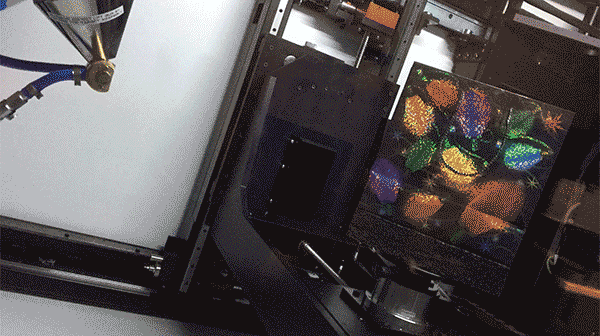
Post by Lola Sanchez-Carrion

Powered by WordPress & Theme by Anders Norén